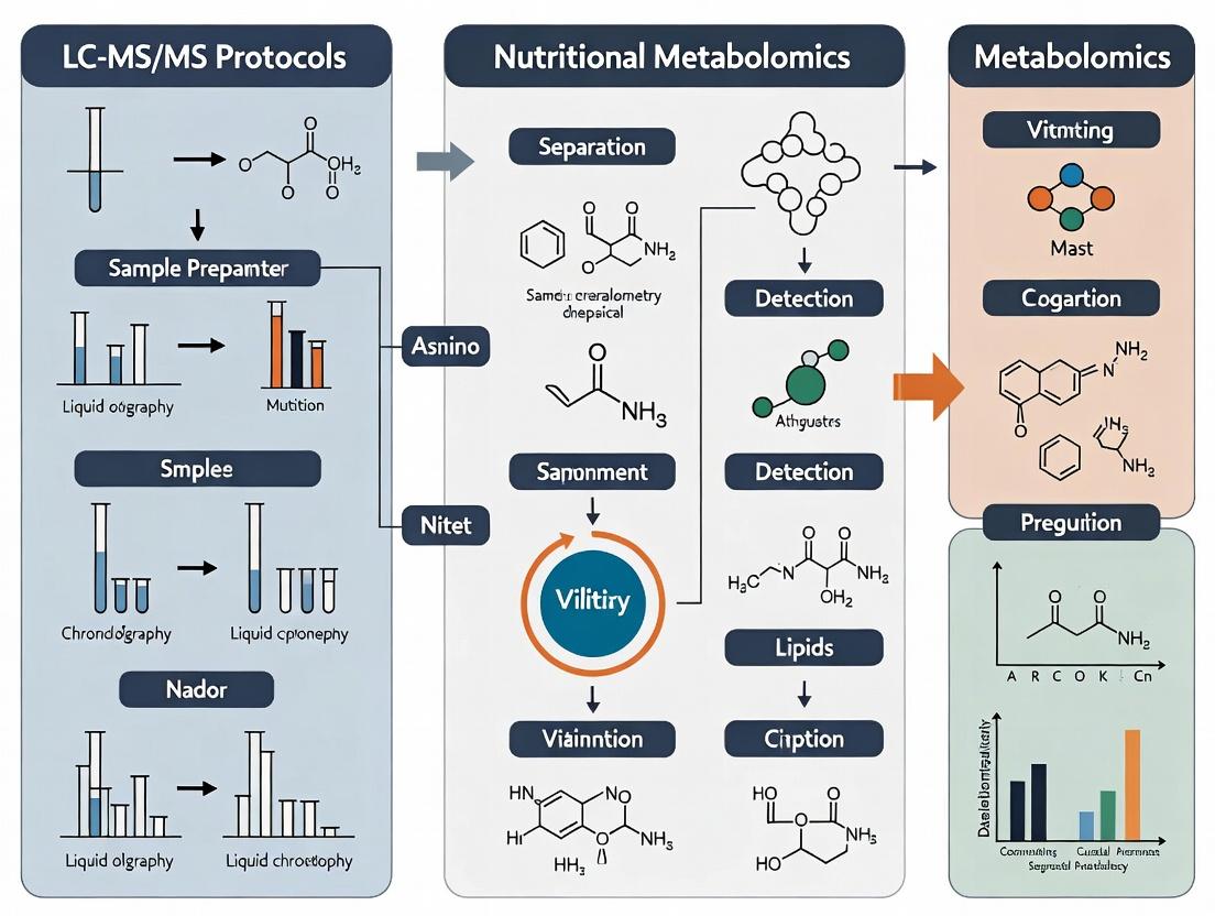
Comprehensive LC-MS/MS Protocols for Nutritional Metabolomics in Human Plasma: From Sample Prep to Biomarker Discovery
This article provides a detailed, step-by-step guide for researchers and scientists implementing LC-MS/MS-based nutritional metabolomics in human plasma.
Comprehensive LC-MS/MS Protocols for Nutritional Metabolomics in Human Plasma: From Sample Prep to Biomarker Discovery
Abstract
This article provides a detailed, step-by-step guide for researchers and scientists implementing LC-MS/MS-based nutritional metabolomics in human plasma. It covers the foundational principles of why plasma is the matrix of choice for assessing dietary intake and nutritional status, followed by comprehensive methodological protocols for sample collection, preparation, chromatography, and tandem mass spectrometry analysis. The guide dedicates significant attention to common troubleshooting scenarios and optimization strategies for sensitivity and reproducibility. Finally, it addresses critical validation parameters, quality control frameworks, and compares LC-MS/MS to alternative analytical platforms. This resource aims to equip professionals in research and drug development with the practical knowledge to generate robust, quantitative metabolomic data for discovering diet-related biomarkers and understanding metabolic pathways.
Nutritional Metabolomics 101: Why Human Plasma is Key to Decoding Diet-Health Interactions
Nutritional metabolomics is defined as the comprehensive profiling of metabolites in biological samples to understand the metabolic response to dietary intake, thereby bridging dietary patterns with measurable metabolic phenotypes. Within human plasma research, LC-MS/MS has become the cornerstone technology due to its high sensitivity, specificity, and ability to quantify a broad spectrum of nutritional biomarkers, from polar vitamins to complex lipids.
Key Applications:
- Biomarker Discovery: Identifying objective biomarkers of food intake (e.g., alkylresorcinols for whole grain, proline betaine for citrus) to move beyond dietary recall inaccuracies.
- Mechanistic Insights: Elucidating metabolic pathways perturbed by specific nutrients or dietary patterns (e.g., saturated fat intake and branched-chain amino acid metabolism).
- Personalized Nutrition: Stratifying individuals based on their metabolic phenotype to predict differential responses to dietary interventions.
- Drug-Nutrient Interactions: Assessing how dietary components modulate drug metabolism pathways and efficacy.
Core Experimental Protocols
Protocol 1: Targeted LC-MS/MS for Quantification of Nutritional Metabolites in Human Plasma
Objective: To precisely quantify a panel of 40 known nutritional biomarkers, including vitamins, carotenoids, and fatty acids.
Materials:
- Sample: EDTA or heparin human plasma (100 µL per analysis).
- Internal Standards: Stable isotope-labeled analogs for each analyte class (e.g., d6-α-Tocopherol, 13C3-carnitine).
- Reagents: HPLC-grade methanol, acetonitrile, water, methyl-tert-butyl ether (MTBE), formic acid, ammonium acetate.
Procedure:
- Sample Preparation (Protein Precipitation & Lipid Extraction):
- Thaw plasma samples on ice.
- Aliquot 100 µL of plasma into a 1.5 mL microcentrifuge tube.
- Add 10 µL of the mixed internal standard working solution.
- Vortex for 10 seconds and incubate for 10 minutes at room temperature.
- Add 300 µL of cold methanol (-20°C) to precipitate proteins. Vortex vigorously for 1 minute.
- For lipid-soluble analytes (e.g., vitamins A, E, carotenoids), add 1 mL of MTBE, vortex for 10 minutes, and centrifuge at 14,000 x g for 10 minutes at 4°C. Transfer the upper organic layer to a new tube and evaporate under nitrogen. Reconstitute in 100 µL of methanol/dichloromethane (1:1).
- For water-soluble analytes, centrifuge the methanol-plasma mixture at 14,000 x g for 15 minutes at 4°C. Transfer the supernatant to a new vial for analysis.
LC-MS/MS Analysis:
- Chromatography: Reversed-phase C18 column (2.1 x 100 mm, 1.7 µm). Mobile Phase A: 0.1% formic acid in water with 5 mM ammonium acetate. Mobile Phase B: 0.1% formic acid in acetonitrile.
- Gradient: 5% B to 95% B over 12 minutes, hold for 3 minutes, re-equilibrate for 5 minutes. Flow rate: 0.35 mL/min. Column temperature: 40°C.
- Mass Spectrometry: Triple quadrupole MS operated in multiple reaction monitoring (MRM) mode. Electrospray ionization (ESI) positive/negative switching. Source parameters: Capillary voltage 3.0 kV, source temperature 150°C, desolvation temperature 500°C, desolvation gas flow 800 L/h.
- Data Acquisition: Optimized MRM transitions, dwell times (≥20 ms), and collision energies for each analyte and its corresponding internal standard are defined in the method table.
Data Processing:
- Quantify using the internal standard method. Generate calibration curves (1-1000 ng/mL) for each analyte using analyte/IS peak area ratios. Apply linear regression with 1/x weighting.
Protocol 2: Untargeted LC-MS/MS for Phenotype Discovery
Objective: To perform global metabolic profiling for hypothesis generation.
Procedure:
- Sample Preparation: As per Protocol 1, but without class-specific extraction. Use a simple methanol precipitation. Pool equal aliquots of all samples to create a quality control (QC) sample.
- LC-MS/MS Analysis: Utilize high-resolution mass spectrometry (e.g., Q-TOF).
- Perform both reversed-phase (C18) and hydrophilic interaction liquid chromatography (HILIC) separations to capture a wider metabolite range.
- Acquire data in data-independent acquisition (DIA) or information-dependent acquisition (IDA) mode with MS/MS fragmentation.
- Data Processing:
- Use software (e.g., MS-DIAL, XCMS) for peak picking, alignment, and compound identification via spectral matching to public libraries (e.g., MassBank, HMDB).
- Normalize data to total ion count or internal standards, then perform multivariate statistical analysis (PCA, PLS-DA) to identify differentially abundant features.
Data Presentation
Table 1: Key Nutritional Biomarkers and Their Analytical Parameters in Human Plasma
| Analyte Class | Example Metabolites | Dietary Source | Typical Plasma Concentration (Fasting) | MRM Transition (Quantifier) | Retention Time (min) | Internal Standard |
|---|---|---|---|---|---|---|
| Carotenoids | β-Carotene, Lutein | Carrots, Leafy greens | 0.1 - 0.8 µmol/L | 537.4 > 445.4 (ESI+) | 8.2 | d6-β-Carotene |
| Fat-Soluble Vitamins | α-Tocopherol (Vit E) | Nuts, Seeds | 15 - 40 µmol/L | 431.4 > 165.1 (ESI+) | 7.5 | d6-α-Tocopherol |
| Water-Soluble Vitamins | Vitamin B5 (Pantothenate) | Meat, Whole grains | 0.5 - 2.0 µmol/L | 220.1 > 90.0 (ESI+) | 2.1 | 13C3-Pantothenate |
| Phytochemicals | Alkylresorcinols C17:0 | Whole grain rye/wheat | 10 - 100 nmol/L | 363.3 > 181.1 (ESI+) | 9.8 | d5-Alkylresorcinol C19:0 |
| Fatty Acids | Omega-3 EPA | Fatty fish | 50 - 150 µmol/L (total) | 301.2 > 257.2 (ESI-) | 6.7 | d5-EPA |
Table 2: Typical LC-MS/MS System Suitability Criteria for Nutritional Metabolomics
| Parameter | Acceptance Criteria | Purpose |
|---|---|---|
| Retention Time Shift | ≤ ± 0.1 min | Chromatographic reproducibility |
| Peak Width (at 50%) | ≤ 0.2 min | Adequate chromatographic resolution |
| Signal/Noise Ratio (Low Calibrator) | ≥ 10:1 | Assay sensitivity |
| QC Sample Precision (CV%) | ≤ 15% (≤ 20% for LLOQ) | Intra-batch reproducibility |
| Calibration Curve R² | ≥ 0.99 | Linearity of response |
Diagrams
Workflow from Diet to Metabolic Phenotype
BCAA Metabolism Links Diet to Insulin Phenotype
The Scientist's Toolkit
| Research Reagent / Solution | Function in Nutritional Metabolomics |
|---|---|
| Stable Isotope-Labeled Internal Standards (e.g., 13C, 15N, d-) | Corrects for matrix effects and losses during sample prep; essential for accurate quantification. |
| Dextran-Coated Charcoal-Stripped Human Plasma | Provides an analyte-free matrix for preparing calibration standards in a matched biological background. |
| SPE Cartridges (Mixed-Mode, C18, HLB) | For selective cleanup and enrichment of specific metabolite classes (e.g., acids, lipids) from plasma. |
| Derivatization Reagents (e.g., Dansyl Chloride) | Enhances detection sensitivity and chromatography of poorly ionizing metabolites (e.g., amines, phenols). |
| Synthetic MRM Standard Kits | Pre-optimized, quantitative standards for targeted panels of nutritional biomarkers (e.g., B vitamins). |
| Quality Control (QC) Plasma Pools | Large-volume, homogeneous pools from representative donors for long-term method performance monitoring. |
| Mass Spectral Libraries (e.g., NIST, HMDB, MoNA) | Critical for annotating unknown peaks in untargeted workflows based on MS/MS fragmentation patterns. |
The Unique Advantages of Human Plasma as a Metabolic Snapshot
Human plasma serves as a comprehensive, systemic biofluid, providing a dynamic snapshot of an individual's metabolic state. It integrates endogenous metabolic pathways, dietary intake, xenobiotic exposure, and gut microbiota activity, making it an ideal matrix for nutritional metabolomics. Within LC-MS/MS-based research, plasma offers unique advantages: standardized collection protocols, rich quantitative data reflective of systemic physiology, and the ability to correlate metabolite shifts with health outcomes. These Application Notes detail the protocols and considerations for leveraging plasma in nutritional metabolomics studies, framed within robust LC-MS/MS workflows.
Plasma, the cell-free fraction of blood, is in constant equilibrium with tissues and organs. It carries nutrients, hormones, signaling molecules, and waste products, offering a real-time, integrated readout of the body's biochemical status. In nutritional metabolomics, this is critical for assessing dietary biomarker discovery, nutrient status, metabolic flexibility, and the physiological response to interventions. Compared to other biofluids like urine or saliva, plasma provides more stable concentrations of a wide range of low-abundance metabolites and is less subject to transient fluctuations.
Key Quantitative Advantages of Human Plasma
The quantitative profile of human plasma provides a rich data source for metabolomic investigation. Key classes of measurable analytes are summarized below.
Table 1: Key Metabolite Classes in Human Plasma Accessible via LC-MS/MS
| Metabolite Class | Example Analytes | Typical Concentration Range | Primary Information Relevance |
|---|---|---|---|
| Amino Acids & Derivatives | Leucine, Isoleucine, Valine, Tryptophan, Kynurenine | 10-500 µM | Protein metabolism, dietary intake, immune regulation |
| Lipids & Fatty Acids | Non-esterified Fatty Acids (NEFA), Lysophosphatidylcholines (LPC), Acylcarnitines | 0.1-1000 µM (class-dependent) | Energy metabolism, membrane integrity, inflammation |
| Carbohydrates & Intermediates | Glucose, Lactate, Citrate, Succinate | 1-5000 µM | Glycolysis, TCA cycle activity, energy state |
| Bile Acids | Cholic acid, Chenodeoxycholic acid, Glyco-conjugates | 0.01-10 µM | Gut microbiome co-metabolism, lipid digestion |
| Vitamins & Cofactors | Vitamin D metabolites, B vitamins (e.g., B12, Folate) | pM to nM | Nutritional status, enzymatic function |
| Xenobiotics | Pharmaceuticals, Food Bioactives (e.g., polyphenols) | Variable | Drug pharmacokinetics, dietary exposure |
Detailed LC-MS/MS Protocol for Broad-Spectrum Plasma Metabolomics
3.1. Materials & Reagent Solutions (The Scientist's Toolkit) Table 2: Essential Research Reagent Solutions
| Item | Function & Critical Notes |
|---|---|
| Ice-cold Methanol (80%, v/v) | Protein precipitation solvent. High purity (LC-MS grade) is essential to minimize background noise. |
| Internal Standard (IS) Mix | A cocktail of stable isotope-labeled analogs (e.g., 13C, 15N) for key metabolite classes. Corrects for extraction efficiency and MS instrument variability. |
| Ammonium Formate / Formic Acid | Common mobile phase additives for positive ion mode LC-MS. Maintains consistent pH and improves ionization. |
| Ammonium Acetate / Ammonium Hydroxide | Common mobile phase additives for negative ion mode LC-MS. |
| C18 & HILIC Chromatography Columns | Complementary separation mechanisms. C18 for lipids, bile acids; HILIC for polar metabolites (amino acids, sugars). |
| Quality Control (QC) Pooled Plasma Sample | Generated by combining small aliquots of all study samples. Injected repeatedly throughout the run to monitor system stability and for data normalization. |
3.2. Pre-Analytical Protocol: Plasma Collection & Metabolite Extraction
- Blood Collection & Processing: Collect venous blood into EDTA or lithium heparin tubes. Critical: Maintain consistent pre-analytical variables (fasting status, time of day, processing time). Process within 30 minutes by centrifugation at 1500-2000 x g for 10-15 minutes at 4°C. Immediately aliquot plasma and store at -80°C.
- Protein Precipitation Extraction:
- Thaw aliquots on ice.
- Pipette 50 µL of plasma into a pre-chilled microcentrifuge tube.
- Add 150 µL of ice-cold 80% methanol containing the internal standard mix.
- Vortex vigorously for 30 seconds.
- Incubate at -20°C for 1 hour to enhance protein precipitation.
- Centrifuge at 17,000 x g for 15 minutes at 4°C.
- Transfer 150 µL of the clear supernatant to a fresh LC-MS vial.
- Dry under a gentle stream of nitrogen or using a vacuum concentrator.
- Reconstitute in 50 µL of a solvent compatible with your initial LC mobile phase (e.g., water or starting mobile phase). Vortex and centrifuge before injection.
3.3. LC-MS/MS Acquisition Parameters (Example)
- Chromatography: Employ two complementary methods.
- Reversed-Phase (C18): For lipids and less polar metabolites. Gradient: Water/Acetonitrile with 0.1% formic acid.
- HILIC: For polar metabolites. Gradient: Acetonitrile/Water with 10mM ammonium acetate (pH 9.0).
- Mass Spectrometry:
- Ionization: Electrospray Ionization (ESI), positive and negative modes.
- Scan Type: Use a combination of Full Scan (m/z 50-1200) for untargeted discovery and Multiple Reaction Monitoring (MRM) for targeted, high-sensitivity quantification of pre-defined panels.
- Resolution: Typically use a triple quadrupole (QqQ) for quantification or a high-resolution (Q-TOF, Orbitrap) for untargeted profiling.
3.4. Data Processing & Analysis
- Use vendor or open-source software (e.g., MS-DIAL, XCMS) for peak picking, alignment, and deconvolution.
- Normalize data using internal standards and QC-based methods (e.g., LOESS, SERRF).
- Perform statistical analysis (univariate t-tests, ANOVA; multivariate PCA, PLS-DA) to identify significant metabolites.
- Annotate significant features using accurate mass, MS/MS spectra, and retention time matching against databases (HMDB, METLIN).
Visualizing Metabolic Pathways & Workflows
Plasma Integrates Systemic Metabolism for LC-MS Analysis
Plasma Metabolite Extraction Protocol for LC-MS
Plasma Reflects Multiple Metabolic Axes
Within nutritional metabolomics, the simultaneous quantification of core compound classes—vitamins, lipids, amino acids, and microbial metabolites—provides a systems-level view of nutritional status, metabolic flux, and host-microbiome interactions. LC-MS/MS is the cornerstone technology due to its specificity, sensitivity, and ability to handle complex matrices like human plasma. This document presents integrated protocols for the targeted analysis of these analytes, framed within a thesis focused on robust LC-MS/MS workflows for human plasma research.
Key Research Reagent Solutions
| Reagent/Material | Function in Analysis |
|---|---|
| Stable Isotope-Labeled Internal Standards (e.g., 13C, 15N, 2H) | Corrects for matrix effects, ionization efficiency variability, and preparation losses for precise quantification. |
| Methanol (LC-MS Grade) | Protein precipitation agent; ensures high-purity, low-background sample cleanup. |
| Acetonitrile (LC-MS Grade) | Mobile phase component; offers different selectivity compared to methanol for chromatographic separation. |
| Ammonium Formate / Formic Acid (MS Grade) | Mobile phase additives for controlling pH and promoting [M+H]+ ionization in positive electrospray mode. |
| Ammonium Acetate / Acetic Acid (MS Grade) | Mobile phase additives for negative ion mode optimization and alternative buffer system. |
| Solid Phase Extraction (SPE) Plates (e.g., C18, Mixed-Mode) | Enable high-throughput, selective cleanup and concentration of analytes from plasma. |
| Derivatization Reagents (e.g., Dansyl Chloride, APTS) | Enhance ionization efficiency and chromatographic separation of poorly ionizing compounds (e.g., some vitamins). |
| Quality Control (QC) Pooled Plasma | Monitors system stability, reproducibility, and data quality throughout analytical batches. |
Experimental Protocols
Protocol 3.1: Sample Preparation for Comprehensive Profiling
Principle: Simultaneous extraction of metabolites across four core classes with maximum recovery and minimal degradation.
Procedure:
- Thawing: Thaw EDTA plasma samples on ice.
- Aliquoting: Transfer 50 µL of plasma into a pre-cooled 1.5 mL microcentrifuge tube.
- Protein Precipitation: Add 200 µL of ice-cold methanol:acetonitrile (50:50, v/v) containing a cocktail of stable isotope-labeled internal standards for all target analyte classes.
- Vortex & Incubate: Vortex vigorously for 30 seconds, then incubate at -20°C for 30 minutes.
- Centrifugation: Centrifuge at 16,000 × g for 15 minutes at 4°C.
- Collection: Transfer 180 µL of the supernatant to a clean LC-MS vial.
- Evaporation & Reconstitution: Evaporate to dryness under a gentle stream of nitrogen. Reconstitute in 50 µL of initial mobile phase (e.g., 98% Water, 2% Acetonitrile, 0.1% Formic Acid).
- Storage: Place vials in autosampler at 4°C until analysis.
Protocol 3.2: LC-MS/MS Analysis with Polarity Switching
Chromatography Conditions:
- Column: C18 reversed-phase column (e.g., 2.1 x 100 mm, 1.7 µm).
- Mobile Phase A: Water with 0.1% formic acid and 5 mM ammonium formate.
- Mobile Phase B: Acetonitrile with 0.1% formic acid.
- Gradient: 2% B (0-1 min), 2-95% B (1-12 min), 95% B (12-14 min), 95-2% B (14-14.1 min), 2% B (14.1-16 min).
- Flow Rate: 0.4 mL/min.
- Column Temp: 40°C.
- Injection Volume: 5 µL.
Mass Spectrometry Conditions:
- Instrument: Triple quadrupole MS with electrospray ionization (ESI).
- Ionization Mode: Fast polarity switching between positive and negative modes within a single run.
- Source Parameters: Capillary Voltage: ±3.0 kV; Source Temp: 150°C; Desolvation Temp: 500°C; Desolvation Gas Flow: 800 L/hr.
- Data Acquisition: Multiple Reaction Monitoring (MRM). Optimized MRM transitions, collision energies, and cone voltages are pre-defined for each analyte and internal standard. Dwell times adjusted to ensure ≥12 points per peak.
Table 1: Representative Analytical Figures of Merit for Core Compound Classes in Plasma
| Compound Class | Example Analytes | Linear Range (ng/mL) | LLOQ (ng/mL) | Intra-day Precision (%RSD) | Inter-day Precision (%RSD) | Recovery (%) |
|---|---|---|---|---|---|---|
| Fat-Soluble Vitamins | Vitamin A (Retinol), 25-OH Vitamin D3, Vitamin E (α-Tocopherol) | 1 - 500 | 0.5 | 3.2 - 5.8 | 5.1 - 8.7 | 92 - 105 |
| Water-Soluble Vitamins | Vitamin B1 (Thiamine), B6 (Pyridoxal), B9 (Folate), B12 (Cobalamin) | 0.1 - 100 | 0.05 | 4.1 - 7.3 | 6.5 - 10.2 | 88 - 102 |
| Amino Acids | Leucine, Tryptophan, Glutamine, Arginine | 50 - 10,000 | 25 | 2.5 - 4.5 | 3.8 - 6.5 | 95 - 108 |
| Complex Lipids | Phosphatidylcholines (PC), Lysophosphatidylcholines (LPC), Ceramides (Cer) | 10 - 5,000 | 5 | 4.5 - 8.5 | 7.0 - 11.5 | 90 - 103 |
| Microbial Metabolites | Short-Chain Fatty Acids (Butyrate), Indole-3-propionic acid, Trimethylamine N-oxide (TMAO) | 0.5 - 200 | 0.25 | 5.5 - 9.0 | 8.2 - 12.4 | 85 - 98 |
Table 2: Polarity and Key MRM Transitions for Select Analytes
| Analyte | Ionization Polarity | Precursor Ion (m/z) > Product Ion (m/z) | Collision Energy (eV) |
|---|---|---|---|
| 25-OH Vitamin D3 | Positive | 401.3 > 159.1 | 18 |
| Folate (5-MTHF) | Positive | 460.1 > 313.1 | 20 |
| Tryptophan | Positive | 205.1 > 146.1 | 16 |
| Butyric Acid | Negative | 87.0 > 43.0 | 10 |
| PC(34:2) | Positive | 758.6 > 184.1 | 32 |
| TMAO | Positive | 76.1 > 58.1 | 18 |
Visualized Workflows and Pathways
Title: Plasma Metabolite Extraction and LC-MS/MS Workflow
Title: Core Metabolite Interactions in Nutritional Status
Within nutritional metabolomics using LC-MS/MS, the study design fundamentally dictates the validity and scope of biological insights. Cohort, intervention, and cross-sectional designs each offer distinct advantages for profiling human plasma metabolomes, guiding hypothesis generation, and establishing causality in diet-disease relationships.
Design Considerations & Quantitative Comparison
Table 1: Comparative Analysis of Primary Study Designs in Nutritional Metabolomics
| Design Parameter | Prospective Cohort Study | Randomized Controlled Trial (RCT) / Intervention | Cross-Sectional Analysis |
|---|---|---|---|
| Primary Aim | Identify temporal relationships & biomarkers of disease risk. | Establish causal effects of a nutritional intervention. | Snapshot of metabolic associations at a single time point. |
| Timeframe | Long-term (years to decades). | Short to medium-term (weeks to months). | Single time point. |
| LC-MS/MS Sampling | Repeated plasma sampling at baseline and pre-defined intervals/follow-ups. | Pre- and post-intervention sampling; often with run-in/washout phases. | Single plasma sample collection. |
| Key Strength | Assesses long-term diet-metabolite-disease trajectories; real-world relevance. | High internal validity; controls for confounding via randomization. | Logistically simple; rapid hypothesis generation. |
| Major Limitation | Costly, time-consuming; residual confounding. | May lack generalizability; ethical/practical limits on interventions. | Cannot infer causality or temporal sequence. |
| Sample Size Typical Range | 500 - 10,000+ participants. | 20 - 100+ participants per arm. | 100 - 1,000+ participants. |
| Primary Statistical Approach | Time-to-event analysis (Cox regression); mixed models for repeated metabolites. | Paired tests (Wilcoxon, t-test); ANOVA for multi-arm studies. | Correlation analysis; linear/logistic regression. |
| Power in Metabolomics | Powered for clinical endpoints, not for full metabolome discovery (often uses nested case-control). | Powered for specific metabolite changes from intervention. | Often underpowered for high-dimensional discovery without validation cohort. |
Detailed Methodological Protocols for LC-MS/MS Metabolomics
Protocol 2.1: Standardized Plasma Collection & Pre-Analytical Processing for All Designs
Objective: Minimize pre-analytical variation in plasma metabolome profiles. Materials: EDTA or heparin tubes, cryovials, refrigerated centrifuge, -80°C freezer. Procedure:
- Fasting Blood Draw: Collect venous blood following a >8h overnight fast. Use consistent time of day across study.
- Immediate Processing: Centrifuge blood samples at 2,000-2,500 x g for 10-15 minutes at 4°C within 2 hours of collection.
- Plasma Aliquoting: Carefully pipette plasma supernatant into pre-labeled cryovials (typically 50-100 µL aliquots) avoiding the buffy coat.
- Snap-Freezing: Flash-freeze aliquots in liquid nitrogen or a dry ice-ethanol bath.
- Storage: Transfer to -80°C freezer within 1 hour. Maintain consistent freezer conditions. Avoid freeze-thaw cycles.
Protocol 2.2: LC-MS/MS Metabolite Extraction from Human Plasma
Objective: Perform a reproducible protein precipitation and metabolite extraction compatible with hydrophilic interaction liquid chromatography (HILIC) and reversed-phase (RP) LC-MS/MS. Research Reagent Solutions:
- Extraction Solvent: Methanol:Acetonitrile:Water (50:30:20, v/v/v) with 0.1% Formic Acid, chilled to -20°C. Function: Denatures proteins, precipitates macromolecules, and extracts a broad range of polar and semi-polar metabolites.
- Internal Standard Mix: Stable isotope-labeled standards (e.g., amino acids, fatty acids, acylcarnitines) in methanol. Function: Corrects for variability during sample preparation, injection, and ionization.
- Quality Control (QC) Pool: An aliquot prepared by combining equal volumes of all study samples. Function: Monitors instrument stability and data reproducibility throughout the analytical sequence.
Procedure:
- Thaw plasma samples on ice.
- Piper 50 µL of plasma into a pre-cooled 1.5 mL microcentrifuge tube.
- Add 200 µL of chilled extraction solvent and 10 µL of internal standard mix.
- Vortex vigorously for 30 seconds.
- Incubate at -20°C for 60 minutes to complete protein precipitation.
- Centrifuge at 18,000 x g for 15 minutes at 4°C.
- Transfer 180 µL of the supernatant to a clean LC-MS vial with insert.
- Keep vials at 4°C in the autosampler (typically 6°C) for analysis. Inject 5-10 µL for analysis.
Protocol 2.3: Analytical Sequence Design for Large Cohort/Cross-Sectional Studies
Objective: Ensure data quality and correct for instrumental drift in high-throughput analyses. Procedure:
- Conditioning: Inject QC pool sample 5-10 times to condition the LC column and stabilize the MS system.
- Randomization: Inject study samples in a randomized order to avoid batch effects correlated with clinical groups.
- QC Frequency: Inject the QC pool sample after every 6-10 study samples.
- Solvent Blanks: Inject a solvent blank (extraction solvent) after every 20-30 samples to monitor carryover.
- Reference Standards: Inject a mixture of authentic chemical standards at the beginning and end of the sequence to confirm metabolite identity and retention time stability.
The Scientist's Toolkit: Essential Research Reagent Solutions
Table 2: Key Reagents & Materials for LC-MS/MS Nutritional Metabolomics
| Item | Function & Rationale |
|---|---|
| Stable Isotope-Labeled Internal Standards (SIL-IS) | Distinguishes analyte signal from background; corrects for matrix effects & extraction efficiency. Essential for quantitative accuracy. |
| Dual LC Column Set (e.g., C18 RP & HILIC) | Provides complementary separation: RP for lipids/bile acids; HILIC for polar metabolites (amino acids, sugars). Maximizes metabolome coverage. |
| Mobile Phase Additives (FA, NH4Ac, NH4OH) | Formic Acid (FA) for positive ion mode; Ammonium Acetate (NH4Ac) for both modes; Ammonium Hydroxide (NH4OH) for negative mode. Modulate ionization efficiency. |
| Commercial Metabolite Libraries (e.g., NIST, HMDB) | Spectral reference libraries for compound annotation based on accurate mass, retention time, and MS/MS fragmentation patterns. |
| Processed QC Pool Sample | Evaluates system stability, batch-to-batch variation, and data reproducibility. Critical for post-acquisition data QC. |
| Standard Reference Material (SRM 1950) | NIST plasma-based metabolomics reference material. Allows inter-laboratory comparison and method benchmarking. |
| Liquid Handling Robot | Automates plasma aliquoting, protein precipitation, and derivatization steps to improve throughput and reduce human error. |
Visualized Workflows & Relationships
Within LC-MS/MS protocols for nutritional metabolomics in human plasma research, pre-analytical variability is a predominant source of error, potentially eclipsing analytical imprecision. The integrity of metabolomic data is fundamentally established during sample collection and initial processing. This document details standardized application notes and protocols for managing fasting status, selecting collection tubes, and implementing stabilization strategies to ensure reproducible and biologically relevant results in nutritional intervention and biomarker discovery studies.
Fasting Status Protocols
Standardized Fasting Protocol for Nutritional Metabolomics
Objective: To minimize dietary confounders and establish a metabolically stable baseline for longitudinal studies.
Protocol:
- Duration: A minimum 10-hour overnight fast is required. Water intake is permitted.
- Participant Instructions: Provide written instructions prohibiting all food, caloric beverages, alcohol, and strenuous exercise for 12 hours prior to phlebotomy. Medications/supplements should be taken as usual unless specified by the study protocol.
- Timing: Schedule collections for morning hours (e.g., 7:00 - 9:00 AM) to mitigate circadian effects.
- Verification: Document fasting duration and compliance via questionnaire at time of draw.
- Post-Collection: After the fasting draw, a standardized nutritional challenge (e.g., mixed meal tolerance test) may be administered for dynamic metabolomic profiling, with timed collections at 30, 60, 120, and 180 minutes.
Impact Assessment: Key Metabolite Sensitivity to Fasting
Table 1: Representative Metabolite Concentration Shifts Post-Prandial
| Metabolite Class | Example Metabolite | Fasting Concentration (Approx.) | Post-Prandial Change (at 2h) | Notes for LC-MS/MS |
|---|---|---|---|---|
| Lipids | Triglycerides | 0.5-1.5 mM | Increase 50-200% | Major confounder; requires strict fasting. |
| Bile Acids | Cholic Acid | Low nM range | Increase 5-10 fold | Rapid kinetics; critical for gut metabolism studies. |
| Amino Acids | Branched-Chain Amino Acids (Leu, Ile, Val) | 100-300 µM | Increase 20-40% | Dietary protein sensitive. Stable after 8h fast. |
| Carbohydrates | Glucose | 4.0-5.5 mM | Increase 20-50% | Stabilizes after 10h fast. |
| Ketone Bodies | β-Hydroxybutyrate | 0.1-0.5 mM | Decrease >50% | Sensitive indicator of fasting status. |
Blood Collection Tube Selection & Protocols
Comparative Evaluation of Collection Tubes
Objective: To select tubes that maximize analyte stability and minimize interference for broad-spectrum metabolomic profiling.
Protocol for Tube Comparison Study:
- Simultaneous Draw: Collect blood via a multi-draw adapter into the following tube types (in randomized order):
- Tube A: Lithium Heparin (Plasma, no gel separator).
- Tube B: EDTA (K2 or K3).
- Tube C: Sodium Citrate.
- Tube D: Serum Clot Activator (with gel separator).
- Processing: Invert all tubes gently 8-10 times. Process within 30 minutes of draw.
- Plasma Tubes (A-C): Centrifuge at 2000 x g for 15 minutes at 4°C.
- Serum Tube (D): Allow to clot for 30 minutes at RT, then centrifuge as above.
- Aliquotting: Carefully pipette supernatant (avoiding gel or buffy coat) into pre-labeled cryovials. Flash-freeze in liquid nitrogen within 1 hour of collection. Store at ≤ -80°C.
- LC-MS/MS Analysis: Batch analyze aliquots for a panel of key nutritional metabolites (e.g., free fatty acids, amino acids, vitamins, oxidative stress markers).
Table 2: Collection Tube Suitability for Nutritional Metabolomics
| Tube Type (Additive) | Primary Mechanism | Key Advantages for LC-MS/MS | Key Disadvantages & Interferences | Recommended Use |
|---|---|---|---|---|
| EDTA (K2/K3) | Chelates Ca²⁺ | Inhibits phospholipases; superior stability for lipids & labile metabolites. Minimal ion suppression. | Can chelate metal ions in MS source; may affect metal-binding analytes. | GOLD STANDARD for broad metabolomics. |
| Lithium Heparin | Activates antithrombin III | No chelator interference in MS. Suitable for trace metal analysis. | Potential for platelet activation, releasing metabolites. Higher phospholipid content. | Acceptable alternative; monitor for platelet-derived artifacts. |
| Serum (Clot Activator) | Promotes clotting | Larger volume yield. Required for some legacy assays. | Clotting releases platelet metabolites (e.g., serotonin). Gel can adsorb lipophilic analytes. | Not recommended for discovery metabolomics; use only if required. |
| Sodium Fluoride/ Oxalate | Glycolysis inhibitor | Stabilizes glucose. | Highly interfering salts; severe ion suppression in MS. | Avoid for global profiling. Use only for dedicated glucose/lactate assays. |
| Citrate | Chelates Ca²⁺ | Used for coagulation studies. | Large dilution factor (3.2-3.8%), diluting metabolites. Interfering citrate peaks in MS. | Not recommended for quantitative metabolite profiling. |
Stabilization & Processing Protocols
Immediate Stabilization Protocol for Labile Metabolites
Objective: To halt enzymatic degradation and chemical oxidation of sensitive metabolite classes (e.g., antioxidants, acyl-carnitines, nucleotides).
Materials: Pre-chilled tubes, ice-water slurry, pre-added stabilization cocktails.
Protocol:
- Draw Blood directly into pre-chilled (4°C) EDTA tubes.
- Immediate Cooling: Place tube in an ice-water slurry (0°C) within 30 seconds of draw.
- Rapid Processing: Centrifuge at 4°C within 30 minutes. For ultra-labile analytes (e.g., glutathione, ATP), consider adding a stabilization cocktail (e.g., 10 µL of 200 mM iodoacetate per mL blood) to the tube immediately after draw.
- Plasma Treatment: For specific pathways, treat plasma immediately after separation:
- For Thiols/Antioxidants: Add 10 µL of 10% (w/v) perchloric acid or 1 M HCl to 100 µL plasma for protein precipitation and stabilization.
- For Choline/ Betaine: None typically needed with rapid freezing.
- Flash-Freezing: Aliquot stabilized plasma into cryovials and submerge in liquid nitrogen for ≥ 2 minutes before transfer to -80°C.
Critical Time-Delay Experiment Protocol
Objective: To quantify metabolite degradation at room temperature and define the maximum allowable processing delay.
Protocol:
- Draw a single blood sample into a pre-chilled EDTA tube.
- Immediately aliquot the whole blood into 5 identical pre-chilled microtubes.
- Time Points: Process each microtube at a different time point after draw: T0 (immediate), T30min, T1h, T2h, T4h. Hold all tubes at room temperature (simulating worst-case scenario) until processing.
- Centrifuge each tube at its designated time point (4°C, 2000 x g, 15 min), aliquot plasma, and flash-freeze.
- Analyze all aliquots in the same LC-MS/MS batch. Plot metabolite abundance vs. time to establish stability thresholds.
Diagrams
Title: Workflow for Fasting and Dynamic Nutritional Challenge Studies
Title: Collection Tube Selection Guide for Nutritional Metabolomics
The Scientist's Toolkit: Essential Research Reagent Solutions
Table 3: Key Materials for Pre-Analytical Stabilization in Metabolomics
| Item | Function & Rationale | Example/Note |
|---|---|---|
| K2EDTA Blood Collection Tubes | Preferred anticoagulant. Chelates calcium to inhibit coagulation and phospholipase activity, preserving lipidome integrity. | 6 mL or 10 mL tubes (e.g., BD Vacutainer #367525). Use gel-free for metabolomics. |
| Enzyme Inhibitor Cocktails | Stabilize ultra-labile metabolites (e.g., glutathione, nucleotides, acyl-CoAs) by halting enzymatic degradation immediately upon draw. | Commercially available (e.g., PPS, Biopreserver) or lab-made (e.g., iodoacetate for thiols). |
| Pre-Chilled Tube Holders / Ice Slurry | Rapid cooling to 0-4°C slows metabolic activity in whole blood prior to processing, critical for accurate snapshot. | Polystyrene foam racks filled with wet ice/water mixture. |
| Cryogenic Vials (Pre-labeled, Screw-top) | For long-term storage of plasma aliquots at ≤ -80°C. Screw-top with O-ring prevents freeze-drying and sample degradation. | Use internally threaded vials (e.g., Corning, Nunc). Avoid aliquot volumes >500 µL to limit freeze-thaw stress. |
| Liquid Nitrogen Dewar or Pre-Chilled (-80°C) Isopropanol Bath | For rapid, uniform flash-freezing of plasma aliquots. Prevents water crystal formation and preserves metabolite stability. | Essential step prior to transfer to -80°C freezer. |
| Low-Binding Pipette Tips & Microtubes | Minimizes adsorption of hydrophobic or protein-scarce metabolites (e.g., eicosanoids, steroids) to plastic surfaces. | Use polypropylene tubes/ tips with polymer additives (e.g., LoBind from Eppendorf). |
| Internal Standard Mix (for Stabilization QC) | Add at the point of plasma separation to monitor and correct for pre-analytical degradation during processing. | Stable-isotope labeled analogs of labile analytes (e.g., d4-choline, 13C6-glutathione). |
Step-by-Step LC-MS/MS Protocol: Sample Preparation, Chromatography, and Mass Spectrometry Analysis
In LC-MS/MS-based nutritional metabolomics, the accurate profiling of small-molecule metabolites from human plasma is paramount. The initial sample preparation step, deproteinization, is critical for removing interfering proteins, minimizing matrix effects, and ensuring instrument longevity. This application note, framed within a broader thesis on robust LC-MS/MS protocols for nutritional metabolomics, provides a comparative analysis of three core deproteinization techniques: classic Protein Precipitation (PPT), optimized Protein Precipitation (PPT) with novel adsorbents, and Supported Liquid Extraction (SLE). We evaluate their efficiency in metabolite recovery, phospholipid removal, and compatibility with downstream untargeted and targeted analyses.
Table 1: Performance Comparison of Deproteinization Techniques
| Parameter | Classic PPT (Acetonitrile) | Optimized PPT (with Zr-SiO₂) | Supported Liquid Extraction (SLE) |
|---|---|---|---|
| Protein Removal Efficiency | >98% | >99% | >99.5% |
| Phospholipid Removal | ~70% | ~95% | ~85% |
| Average Metabolite Recovery (Polar) | 85-95% | 90-98% | 75-85% |
| Average Metabolite Recovery (Lipophilic) | 60-75% | 85-95% | 90-98% |
| Processed Sample Cleanliness | Moderate | High | High |
| Susceptibility to Matrix Effects (ESI+) | High | Moderate-Low | Low |
| Sample Throughput (96 samples) | ~60 minutes | ~75 minutes | ~90 minutes |
| Organic Solvent Consumption | 300 µL (per 100 µL plasma) | 300 µL (per 100 µL plasma) | 800 µL (per 100 µL plasma) |
| Cost per Sample | Low | Moderate | High |
| Automation Compatibility | Moderate | Moderate | High |
Table 2: Recoveries of Key Metabolite Classes (Mean % ± RSD, n=6)
| Metabolite Class | Example Analyte | Classic PPT | Optimized PPT | SLE |
|---|---|---|---|---|
| Amino Acids | Leucine | 92 ± 4% | 96 ± 3% | 80 ± 6% |
| Carboxylic Acids | Citrate | 88 ± 5% | 94 ± 2% | 78 ± 5% |
| Vitamins | Vitamin B3 (Niacin) | 85 ± 7% | 90 ± 4% | 72 ± 8% |
| Lipids (FFA) | Palmitic Acid | 68 ± 9% | 92 ± 3% | 95 ± 2% |
| Phospholipids | PC(34:2) | 30% Remaining | 5% Remaining | 15% Remaining |
| Steroids | Cortisol | 72 ± 8% | 89 ± 4% | 97 ± 2% |
Detailed Experimental Protocols
Protocol 1: Classic Protein Precipitation (PPT)
- Objective: Rapid, cost-effective protein removal for broad-spectrum metabolomics.
- Materials: Human plasma (100 µL), ice-cold LC-MS grade acetonitrile (300 µL), vortex mixer, microcentrifuge (capable of 13,000 x g), 1.5 mL polypropylene microtubes.
- Procedure:
- Aliquot 100 µL of thawed plasma into a 1.5 mL microtube.
- Add 300 µL of ice-cold acetonitrile.
- Vortex vigorously for 60 seconds.
- Incubate at -20°C for 10 minutes to enhance protein aggregation.
- Centrifuge at 13,000 x g for 10 minutes at 4°C.
- Carefully transfer the clear supernatant (~350 µL) to a clean vial.
- Evaporate to dryness under a gentle stream of nitrogen at 40°C.
- Reconstitute the dried extract in 100 µL of initial LC-MS mobile phase (e.g., 95:5 water:acetonitrile + 0.1% formic acid). Vortex for 60 seconds and centrifuge before LC-MS/MS analysis.
Protocol 2: Optimized PPT with Phospholipid Removal Adsorbent
- Objective: Enhanced phospholipid removal to reduce ion suppression in ESI+ mode.
- Materials: Human plasma (100 µL), ice-cold LC-MS grade acetonitrile (300 µL), zirconia-coated silica (Zr-SiO₂) particles (e.g., 5 mg), vortex mixer, microcentrifuge, 1.5 mL microtubes.
- Procedure:
- Follow steps 1-3 of Protocol 1 (plasma + acetonitrile).
- Add 5 mg of Zr-SiO₂ adsorbent directly to the plasma-acetonitrile mixture.
- Vortex for 120 seconds to ensure complete adsorption of phospholipids.
- Incubate at -20°C for 10 minutes.
- Centrifuge at 13,000 x g for 10 minutes at 4°C.
- Transfer the supernatant, avoiding the pellet of proteins and adsorbent.
- Proceed with evaporation and reconstitution as in Protocol 1 (steps 7-8).
Protocol 3: Supported Liquid Extraction (SLE)
- Objective: High-efficiency, reproducible extraction with superior cleanliness for lipophilic metabolites.
- Materials: Human plasma (100 µL), LC-MS grade water (200 µL), equilibration solution (5% NH₄OH in water, v/v), elution solvent (e.g., methyl tert-butyl ether (MTBE):ethyl acetate, 1:1, v/v), 96-well SLE plate (diatomaceous earth), positive pressure manifold or centrifuge, collection plates, evaporator.
- Procedure:
- Dilute 100 µL plasma with 200 µL of LC-MS grade water and mix gently.
- Condition the SLE plate wells by slowly adding 500 µL of equilibration solution (5% NH₄OH). Allow to soak for 5 minutes, then apply positive pressure/centrifuge to empty.
- Load the diluted plasma sample onto the conditioned SLE bed. Allow it to absorb into the support for 5-10 minutes without airflow.
- Elute metabolites by slowly adding 2 x 400 µL of the organic elution solvent (MTBE:ethyl acetate). Apply gentle pressure/centrifuge after a 2-minute equilibration period for each elution into a clean collection plate.
- Combine the eluates (~800 µL total) and evaporate to complete dryness under nitrogen at 40°C.
- Reconstitute in an appropriate LC-MS compatible solvent (e.g., 80:20 methanol:water) and proceed to analysis.
Visualizations
Plasma Deproteinization Workflow Comparison
Decision Logic for Technique Selection
The Scientist's Toolkit: Essential Research Reagent Solutions
| Item / Reagent Solution | Function & Rationale |
|---|---|
| LC-MS Grade Acetonitrile/Methanol | High-purity, low-UV absorbing solvents for precipitation and reconstitution to prevent background interference in MS detection. |
| Zirconia-Coated Silica (Zr-SiO₂) Particles | Selective adsorbent used in optimized PPT to chelate and remove phospholipids via Lewis acid-base interaction, reducing ion suppression. |
| Supported Liquid Extraction (SLE) Plate | Diatomaceous earth-based support that holds aqueous sample for liquid-liquid extraction with organic solvent, offering reproducibility and automation. |
| Phospholipid Removal SPE Cartridges (e.g., HybridSPE-PPT) | Alternative single-use cartridges combining precipitation and selective filtration for phospholipid removal. |
| Internal Standard Mix (Stable Isotope Labeled) | Added prior to extraction to correct for variability in recovery, evaporation, and matrix effects; critical for quantitative accuracy. |
| MTBE (Methyl tert-butyl ether) | Organic elution solvent for SLE; provides excellent recovery of lipophilic metabolites with low water miscibility. |
| Positive Pressure Manifold (96-well) | Enables simultaneous, controlled processing of multiple SLE or SPE samples, improving throughput and reproducibility over manual methods. |
| Nitrogen Evaporator with Heating Block | For rapid, uniform concentration of organic extracts post-extraction without sample degradation, prior to LC-MS reconstitution. |
Within the framework of a comprehensive thesis on LC-MS/MS protocols for nutritional metabolomics in human plasma, the choice of sample preparation is a critical first step that dictates the breadth and depth of metabolite observation. The fundamental dichotomy lies between targeted extraction, optimized for specific analyte classes, and untargeted extraction, which aims for maximal coverage of the metabolome. The solvent system is the primary lever controlling this selectivity. This application note details contemporary protocols and solvent formulations, balancing extraction efficiency, protein removal, and compatibility with reversed-phase (RP) and hydrophilic interaction liquid chromatography (HILIC) LC-MS/MS analyses.
Core Solvent Systems: Mechanisms and Applications
The efficacy of a solvent system is determined by its ability to precipitate proteins while concurrently solubilizing a wide range of metabolites with varying polarities.
Key Mechanisms:
- Protein Denaturation & Precipitation: Organic solvents (MeOH, ACN) and strong acids (e.g., TCA) disrupt protein hydration shells and charge stability, causing coagulation and removal via centrifugation. This is essential to prevent column fouling and ion suppression.
- Metabolite Solubilization: A biphasic or monophasic mixture determines which metabolite classes partition into the supernatant. Methanol excels at extracting polar and semi-polar metabolites, while acetonitrile offers cleaner backgrounds. Mixtures with water and sometimes chloroform or MTBE enable broader polarity coverage.
Table 1: Quantitative Comparison of Common Solvent Systems for Plasma Metabolomics
| Solvent System (Ratio) | Protein Recovery (%) | Metabolite Class Bias | LC-MS Mode Suitability | Key Advantage | Key Disadvantage |
|---|---|---|---|---|---|
| Methanol (MeOH) 100% | ~95% (Precipitated) | Polar, Semi-polar | RP, HILIC | Excellent for polar central carbon metabolism intermediates; simple protocol. | Poor recovery of very lipophilic metabolites (e.g., triglycerides). |
| Acetonitrile (ACN) 100% | ~98% (Precipitated) | Polar, Semi-polar | RP (optimal) | Superior for RP; less background, sharper peaks. | Can co-precipitate some moderately polar metabolites. |
| MeOH:ACN:H₂O (2:2:1, v/v/v) | ~99% (Precipitated) | Broadest Polar/Semi-polar | RP & HILIC | Combines strengths of MeOH and ACN; minimizes bias; works for lipidomics if biphasic. | Evaporation step required if phase separation occurs. |
| Chloroform:MeOH:H₂O (1:3:1, Bligh & Dyer) | Phase Separates | Lipids (org), Polar (aq) | RP (Lipids) & HILIC (Polar) | True biphasic extraction for simultaneous polar/lipid profiling. | Use of chlorinated solvents; more complex handling. |
| MTBE:MeOH:H₂O (10:3:2.5, Matyash) | Phase Separates | Lipids (org), Polar (aq) | RP (Lipids) & HILIC (Polar) | Less toxic than chloroform; high lipid recovery. | Requires careful phase separation. |
| Trichloroacetic Acid (TCA) 1-5% | ~100% (Precipitated) | Acid-stable Polar (e.g., organic acids) | RP Ion-pairing, HILIC | Very efficient protein removal; good for acidic metabolites. | Harsh acid can degrade labile metabolites (e.g., ATP, some vitamins). |
Detailed Experimental Protocols
Protocol 3.1: Untargeted Extraction Using Modified MeOH:ACN:H₂O
Objective: Maximize coverage of polar to semi-polar metabolites for global profiling. Materials: Human plasma (deproteinized), -80°C storage; LC-MS grade Methanol, Acetonitrile, Water; 1.5 mL microcentrifuge tubes; vacuum concentrator; ultrasonic bath; centrifuge (capable of 14,000 g at 4°C).
- Thawing: Thaw plasma samples on ice.
- Aliquot: Transfer 50 µL of plasma into a pre-chilled 1.5 mL microcentrifuge tube.
- Precipitation/Extraction: Add 200 µL of pre-chilled (-20°C) extraction solvent (Methanol:Acetonitrile:Water, 2:2:1 v/v/v). Vortex vigorously for 30 seconds.
- Incubation: Sonicate in an ice-water bath for 10 minutes.
- Centrifugation: Centrifuge at 14,000 g for 15 minutes at 4°C.
- Collection: Carefully transfer 180 µL of the supernatant (avoiding the protein pellet) to a new, labeled LC-MS vial.
- Drying (Optional): Evaporate to dryness under vacuum and reconstitute in 50 µL of initial LC mobile phase (e.g., 98% H₂O, 2% ACN for HILIC; or 5% ACN, 95% H₂O for RP). Vortex thoroughly. If not drying, dilute 1:1 with aqueous phase.
- Storage: Store at -80°C until LC-MS/MS analysis.
Protocol 3.2: Targeted Lipid Extraction (MTBE Method)
Objective: Selective extraction of lipid classes for targeted lipidomics. Materials: Human plasma; LC-MS grade MTBE, Methanol, Water; 2 mL microcentrifuge tubes; centrifuge.
- Aliquot: Add 50 µL plasma to a 2 mL tube.
- Acidification: Add 150 µL of methanol (containing internal standards). Vortex 10 sec.
- Lipid Extraction: Add 500 µL of Methyl-tert-butyl ether (MTBE). Vortex vigorously for 1 hour at room temperature or shake for 10 min.
- Phase Induction: Add 125 µL of LC-MS grade water to induce phase separation. Vortex for 20 seconds.
- Centrifugation: Centrifuge at 14,000 g for 10 minutes at 20°C.
- Collection: Two clear phases form. The upper (organic) phase contains lipids. Collect ~400 µL of the upper phase into a clean vial.
- Drying: Evaporate under a gentle stream of nitrogen or vacuum.
- Reconstitution: Reconstitute in 100 µL of appropriate solvent (e.g., 9:1 IPA:MeOH or MeOH:CHCl₃). Vortex and sonicate to dissolve.
- Storage: Store at -80°C until analysis.
Visualizations
Title: Solvent System Selection Logic Flow
Title: Generic Plasma Metabolite Extraction Workflow
The Scientist's Toolkit: Research Reagent Solutions
| Item | Function & Rationale |
|---|---|
| LC-MS Grade Solvents (MeOH, ACN, Water, IPA) | Ultra-pure to minimize background chemical noise, ion suppression, and column contamination. Essential for sensitive detection. |
| Methyl-tert-butyl ether (MTBE) | A less toxic, less dense alternative to chloroform for biphasic lipid extractions. Forms a distinct upper organic phase. |
| Internal Standard Mixtures | A cocktail of isotopically labeled analogs (e.g., ¹³C, ²H) of various metabolite classes. Spiked-in pre-extraction to correct for losses during preparation and matrix effects during MS analysis. |
| Protein LoBind Tubes (1.5-2 mL) | Minimize non-specific adsorption of metabolites (especially lipids and peptides) to tube walls, improving recovery and reproducibility. |
| Vacuum Concentrator (SpeedVac) | For gentle, uniform removal of extraction solvents without excessive heat, prior to reconstitution in LC-compatible buffer. |
| C18 & HybridSPE-Precipitation Plates | For automated, high-throughput protein precipitation. HybridSPE uses zirconia-coated silica to selectively remove phospholipids—a major source of ion suppression. |
| Biological pH Buffers (e.g., Ammonium Acetate, Formate) | Used in extraction or reconstitution to stabilize pH-sensitive metabolites and enhance ionization efficiency in ESI-MS. |
Within the framework of a thesis on LC-MS/MS protocols for human plasma nutritional metabolomics, column chemistry selection is a pivotal initial step. Human plasma contains a highly complex mixture of metabolites spanning a wide polarity range, from polar amino acids and sugars to non-polar lipids and fat-soluble vitamins. The choice between Hydrophilic Interaction Liquid Chromatography (HILIC) and Reversed-Phase (RP) chromatography dictates metabolite coverage, sensitivity, and overall analytical success.
Principle and Application Comparison
HILIC employs a polar stationary phase (e.g., bare silica, amino, amide) with a hydrophobic mobile phase (high organic, e.g., acetonitrile). Analytes elute in order of increasing polarity. It is ideal for retaining and separating small, polar, and ionic compounds that elute too quickly or not at all in RP.
Reversed-Phase utilizes a non-polar stationary phase (e.g., C18, C8) with a polar mobile phase (water/organic gradient). Analytes elute in order of decreasing polarity. It is the gold standard for medium to non-polar compounds.
Table 1: Direct Comparison of HILIC vs. Reversed-Phase for Metabolomics
| Parameter | HILIC | Reversed-Phase (C18) |
|---|---|---|
| Stationary Phase | Polar (Silica, Amide, Diol) | Non-polar (C18, C8, Phenyl) |
| Mobile Phase Start | High Organic (≥70% ACN) | High Aqueous (≥90% Water) |
| Elution Order | Polar Last | Polar First |
| Optimal for | Polar metabolites (Sugars, Organic acids, Nucleotides, Polar lipids) | Mid-to-Non-polar metabolites (Lipids, Steroids, Fat-soluble vitamins, Flavonoids) |
| Plasma Sample Prep | Protein precipitation with ACN recommended; supernatant compatible with injection. | Protein precipitation with MeOH or ACN; may require evaporation/reconstitution. |
| Gradient Time (Typical) | 15-25 min | 15-30 min |
| MS Compatibility | High initial organic can enhance ionization. | May require post-column addition for optimal ionization of early eluters. |
| Key Challenge | Long column equilibration, sensitivity to buffer conc. | Poor retention of very polar metabolites. |
Detailed Experimental Protocols
Protocol 3.1: Two-Platform Metabolomics Screening for Human Plasma
Objective: To comprehensively cover a broad metabolite spectrum from a single plasma extract using complementary HILIC and RP separations coupled to high-resolution MS/MS.
Materials & Reagents:
- Human plasma samples (post-prandial, fasted state).
- Cold HPLC-grade methanol, acetonitrile (ACN), and water.
- Ammonium acetate, formic acid (MS grade).
- Internal Standard Mix: Stable isotope-labeled amino acids, fatty acids, sugars (e.g., Cambridge Isotope Laboratories).
- HILIC Column: e.g., Acquity UPLC BEH Amide (2.1 x 100 mm, 1.7 µm).
- RP Column: e.g., Acquity UPLC HSS T3 (2.1 x 100 mm, 1.8 µm).
Procedure:
- Sample Preparation:
- Thaw plasma on ice. Vortex briefly.
- Aliquot 50 µL plasma into a microcentrifuge tube.
- Add 150 µL of cold MeOH:ACN (1:1, v/v) containing the internal standard mix.
- Vortex vigorously for 1 min. Incubate at -20°C for 1 hour to precipitate proteins.
- Centrifuge at 17,000 x g for 15 min at 4°C.
- Transfer 150 µL of supernatant to a fresh LC vial for RP analysis.
- For HILIC analysis, take a separate 50 µL aliquot of plasma, precipitate with 200 µL of cold ACN, centrifuge, and use the supernatant directly.
LC-MS/MS Conditions (RP):
- Column: HSS T3, 45°C.
- Mobile Phase: A: 0.1% Formic acid in water; B: 0.1% Formic acid in ACN.
- Gradient: 1% B to 99% B over 18 min, hold 2 min, re-equilibrate for 5 min.
- Flow Rate: 0.4 mL/min. Injection: 2 µL.
- MS: ESI positive/negative switching, data-dependent acquisition (DDA).
LC-MS/MS Conditions (HILIC):
- Column: BEH Amide, 45°C.
- Mobile Phase: A: 10mM Ammonium acetate in 95% ACN, pH 9.0; B: 10mM Ammonium acetate in water, pH 9.0.
- Gradient: 95% A to 60% A over 16 min, hold 2 min, re-equilibrate at 95% A for 7 min.
- Flow Rate: 0.5 mL/min. Injection: 3 µL.
- MS: ESI positive/negative switching, DDA.
Protocol 3.2: Targeted Bile Acid Analysis via HILIC
Objective: Separate and quantify isomeric conjugated bile acids (polar) in human plasma.
Procedure:
- Sample Prep: As per HILIC prep above (ACN precipitation).
- LC Conditions:
- Column: ZIC-pHILIC (150 x 2.1 mm, 5 µm).
- Mobile Phase: A: 20mM Ammonium carbonate in water; B: ACN.
- Gradient: 80% B to 50% B over 12 min.
- Flow: 0.2 mL/min. Temp: 40°C. Inj: 5 µL.
- MS/MS: Negative ESI, Multiple Reaction Monitoring (MRM).
Protocol 3.3: Comprehensive Lipidomics via Reversed-Phase
Objective: Profile non-polar to mid-polar lipid classes (TAG, DAG, PL, CE) in a single run. Procedure:
- Sample Prep: Use RP preparation (MeOH:ACN).
- LC Conditions:
- Column: CORTECS C18+ (2.1 x 100 mm, 1.6 µm).
- Mobile Phase: A: 10mM Ammonium formate in 60% ACN/Water; B: 10mM Ammonium formate in 90% IPA/ACN.
- Gradient: 40% B to 100% B over 20 min.
- Flow: 0.4 mL/min. Temp: 55°C.
- MS/MS: Positive/Negative ESI with polarity switching.
Visualizations
Diagram Title: LC Column Selection Workflow for Metabolites
Diagram Title: Dual-Platform Metabolomics Analysis Protocol
The Scientist's Toolkit: Key Research Reagent Solutions
Table 2: Essential Materials for LC-MS/MS Metabolomics of Plasma
| Item | Function & Rationale |
|---|---|
| HILIC Column (e.g., BEH Amide) | Provides strong retention of polar metabolites via hydrogen bonding and dipole-dipole interactions. Critical for analyzing sugars, organic acids, and polar lipids. |
| RP Column (e.g., C18 with aqueous retention) | Standard workhorse for lipidomics and mid-polar metabolites. Modern phases like T3 or C18+ offer better retention for some polar compounds. |
| Stable Isotope Internal Standards (SIL-IS) | Corrects for matrix effects and extraction variability. Essential for accurate quantification. Include a mix spanning polar/non-polar classes. |
| MS-Grade Ammonium Salts (Acetate/Formate) | Provides volatile buffering for mobile phases. Ammonium acetate (pH~9) is common for HILIC; formate is common for RP in positive ESI. |
| Cold Methanol & Acetonitrile (HPLC Grade) | Solvents for protein precipitation. ACN is preferred for HILIC-friendly extracts. MeOH is more efficient for broad precipitation. |
| Solid Phase Extraction (SPE) Plates (C18, Mixed-Mode) | For advanced sample clean-up to remove phospholipids and salts, reducing ion suppression and column contamination. |
Within the broader thesis on developing robust LC-MS/MS protocols for nutritional metabolomics in human plasma research, mobile phase optimization is a critical pillar. The complexity of the plasma metabolome—spanning polar vitamins, hydrophobic lipids, and ionic organic acids—demands meticulous attention to chromatographic conditions. Poor peak shape, manifested as fronting, tailing, or broadening, directly compromises sensitivity, reproducibility, and accurate quantification, ultimately jeopardizing the integrity of nutritional intervention studies. This application note details systematic strategies for optimizing aqueous and organic mobile phases through buffer selection, additive use, and gradient profile design to achieve sharp, symmetric peaks for diverse metabolite classes.
Core Principles & Recent Findings
Live search data confirms that current best practices emphasize volatile buffers compatible with MS detection. Key trends include:
- Ammonium Formate/Acetate Dominance: These remain the gold-standard buffers (typically 2-10 mM, pH 3.5-5.0) for positive- and negative-mode electrospray ionization (ESI), respectively.
- Additives for Challenging Analytes: For acidic metabolites (e.g., short-chain fatty acids, bile acids), 0.1% formic acid is standard for positive mode, while low concentrations (0.1-1 mM) of hydrophilic ion-pairing agents like dimethylhexylamine (DMHA) can improve peak shape for polar anions without severe MS suppression. Conversely, for basic compounds, amines like diethylamine (DEA, 0.01-0.1%) can mitigate silanol interactions.
- Gradient Steepness and Re-equilibration: A linear gradient with a slope of 1-5% organic modifier per minute is often optimal for complex metabolomic samples. Adequate column re-equilibration (>5 column volumes) between runs is non-negotiable for retention time stability.
- Column Chemistry Synergy: Optimization is inseparable from stationary phase choice. For instance, charged surface hybrid (CSH) columns benefit from specific additive adjustments.
Key Research Reagent Solutions
The following table lists essential materials for mobile phase optimization in nutritional metabolomics LC-MS/MS.
Table 1: Essential Research Reagents for Mobile Phase Optimization
| Reagent/ Material | Function in Mobile Phase Optimization |
|---|---|
| Ammonium Formate (LC-MS Grade) | Volatile buffer salt for pH control in positive and negative ESI; reduces adduct formation. |
| Ammonium Acetate (LC-MS Grade) | Volatile buffer for pH control; often preferred in negative ESI for some analyte classes. |
| Formic Acid (LC-MS Grade, ≥99%) | Common acidic additive (0.05-0.1%) to promote protonation in positive ESI and improve peak shape for acids. |
| Acetic Acid (LC-MS Grade) | Milder acidic alternative to formic acid; sometimes reduces in-source fragmentation. |
| Ammonium Hydroxide (LC-MS Grade) | Basic additive for negative ESI to promote deprotonation; used sparingly to adjust pH. |
| Dimethylhexylamine (DMHA) | Hydrophilic ion-pairing agent at low concentration (e.g., 1 mM) to improve peak shape of polar anions (TCA cycle intermediates). |
| Diethylamine (DEA) | Basic additive (0.01-0.1%) to block active silanol sites and reduce tailing of basic metabolites. |
| Water (LC-MS Grade) | Aqueous mobile phase base; must be ultra-pure, organic-free. |
| Acetonitrile (LC-MS Grade) | Primary organic modifier; provides low viscosity and background, high elution strength. |
| Methanol (LC-MS Grade) | Alternative organic modifier; different selectivity for some lipid classes; higher viscosity. |
Experimental Protocols
Protocol 1: Systematic Screening of Buffer pH and Additives
Objective: Identify optimal pH and additive for peak symmetry of target metabolite panels.
- Prepare Stock Buffers: Prepare 100 mM ammonium formate (for pH 3.0-5.0) and ammonium acetate (for pH 5.0-8.0) solutions in LC-MS grade water. Adjust pH with formic acid or ammonium hydroxide as needed.
- Prepare Mobile Phases: Create a matrix of mobile phase A solutions: 5 mM buffer at pH 3.0, 4.0, 5.0, 6.0, 7.0, and 8.0, each with and without 0.1% formic acid (for positive mode) or 0.1% ammonium hydroxide (for negative mode). Mobile phase B is acetonitrile with same additive as paired mobile phase A.
- Chromatography: Inject a standardized plasma metabolite extract containing representative acids, bases, and neutrals. Use a shallow, generic gradient (e.g., 5-95% B in 10 min) on a C18 column.
- Analysis: Calculate asymmetry factor (As) at 10% peak height for each analyte. Select condition yielding As closest to 1.0 for the broadest set of critical analytes.
Protocol 2: Optimizing Gradient Elution Profile
Objective: Develop a gradient that maximizes peak capacity and resolution for a wide metabolomic scope.
- Initial Scouting Run: Using the best buffer/additive from Protocol 1, perform a fast gradient from 1% to 99% B in 15 minutes.
- Analyze Distribution: Plot detected peaks by their retention time and logP (if known). Identify regions of co-elution and excessive void time.
- Design Multi-Segment Gradient: Introduce a shallow initial segment (e.g., 1-10% B over 3 min) to resolve polar metabolites. Steepen the gradient for mid-hydrophobicity compounds (10-70% B over 8 min), and use a final sharp rise for lipids (70-99% B in 2 min).
- Validate and Adjust: Inject the sample with the new gradient. Adjust segment slopes to evenly distribute peaks. Ensure a 5-minute re-equilibration at initial conditions.
Protocol 3: Evaluating Ion-Pairing Additives for Polar Anions
Objective: Improve peak shape and retention of TCA cycle intermediates and other carboxylic acids.
- Prepare Additive Solutions: Prepare mobile phase A (water) with: a) 0.1% formic acid (control), b) 10 mM ammonium acetate pH 6.5, c) 10 mM ammonium acetate pH 6.5 + 1 mM DMHA.
- Chromatography: Use a HILIC or reversed-phase column. Inject a standard mix of polar acids (e.g., citrate, succinate, malate, α-ketoglutarate). Use an isocratic or shallow gradient method.
- Assessment: Compare peak shape, retention factor (k), and signal intensity. Note: DMHA requires extensive column washing (≥30 column volumes) to remove.
Data Presentation
Table 2: Effect of Buffer pH and Additive on Peak Asymmetry (As) of Representative Metabolites (C18 Column, Positive ESI)
| Metabolite Class | Example | 5 mM NH₄Frm, pH 3.0 | 5 mM NH₄Frm, pH 3.0 + 0.1% FA | 5 mM NH₄Frm, pH 5.0 | 5 mM NH₄Frm, pH 5.0 + 0.1% FA |
|---|---|---|---|---|---|
| Basic | Choline | 1.85 (Tailing) | 1.15 | 2.10 | 1.25 |
| Acidic | Pantothenate | 0.92 | 0.95 | 1.05 | 1.08 |
| Neutral | Glucose | 1.10 | 1.12 | 1.08 | 1.10 |
| Amphoteric | Tryptophan | 1.02 | 1.01 | 1.20 | 1.18 |
Table 3: Impact of Gradient Slope on Peak Parameters in Plasma Metabolomics
| Gradient Slope (%B/min) | Average Peak Width (min) | Average Asymmetry Factor | Number of Peaks Detected (m/z 50-1000) |
|---|---|---|---|
| 2 | 0.18 | 1.05 | 450 |
| 5 | 0.22 | 1.08 | 425 |
| 10 | 0.35 | 1.15 | 380 |
Visualized Workflows & Relationships
Title: Mobile Phase Optimization Decision Workflow
Title: Gradient Steepness Effect on Chromatographic Peaks
Within the framework of LC-MS/MS protocols for nutritional metabolomics in human plasma research, the selection of detection mode is paramount. This application note details two core MS/MS strategies: Multiple Reaction Monitoring (MRM) for targeted quantification and Data-Dependent Acquisition (DDA) for untargeted discovery. The former delivers high sensitivity and precision for known metabolites, while the latter enables hypothesis-free profiling of the plasma metabolome to identify novel nutritional biomarkers.
Core Detection Modes: Principles and Applications
Targeted Quantification via Multiple Reaction Monitoring (MRM)
MRM on triple quadrupole mass spectrometers is the gold standard for quantifying predefined metabolites with high accuracy, precision, and sensitivity. It is ideal for validating hypotheses, conducting large cohort studies, and performing absolute quantification.
Principle: The first quadrupole (Q1) filters for the precursor ion of a specific metabolite. The second quadrupole (q2, collision cell) fragments the ion. The third quadrupole (Q3) filters for a unique, abundant product ion. This two-stage filtering drastically reduces chemical noise.
Key Applications in Nutritional Metabolomics:
- Quantification of vitamin metabolites (e.g., A, D, B9, B12).
- Measurement of essential fatty acids and their oxidation products.
- Monitoring of amino acid levels post-intervention.
- Validation of candidate biomarkers from discovery-phase studies.
Untargeted Discovery via Data-Dependent Acquisition (DDA)
DDA, typically on quadrupole-time-of-flight (Q-TOF) or Orbitrap instruments, is used for comprehensive profiling of the metabolome without prior target lists. It is essential for discovery-phase research to identify differentially expressed metabolites in response to dietary interventions.
Principle: The instrument performs an initial MS1 scan to record all ions within a mass range. In real-time, it selects the most intense (or other predefined criteria) precursor ions from the MS1 scan for subsequent MS2 fragmentation. This cycle repeats, building a library of fragmentation spectra for compound identification.
Key Applications in Nutritional Metabolomics:
- Discovery of novel dietary biomarkers.
- Profiling of unknown metabolic shifts.
- Generating hypotheses on metabolic pathways affected by nutrition.
- Compound identification in complex plasma samples.
Table 1: Comparative Overview of MRM and DDA Modes
| Feature | MRM (Targeted Quantification) | DDA (Untargeted Discovery) |
|---|---|---|
| Primary Goal | High-quality quantification of known analytes. | Identification of unknown or unexpected compounds. |
| Instrument Type | Triple Quadrupole (QqQ). | Q-TOF, Quadrupole-Orbitrap. |
| Throughput | High (10s-100s of targets per method). | Moderate (limited by cycle time). |
| Sensitivity | Excellent (fg-pg on-column). | Good (pg-ng on-column). |
| Dynamic Range | 4-6 orders of magnitude. | 3-4 orders of magnitude. |
| Quantitative Rigor | Excellent (uses internal standards). | Semi-quantitative (relative comparison). |
| Identification Power | Low (confirmation only). | High (MS/MS spectra for library matching). |
| Optimal Use Case | Validating & quantifying predefined panels (e.g., vitamins, bile acids). | Discovery of novel biomarkers, pathway analysis. |
Detailed Experimental Protocols
Protocol 1: MRM Quantification of Fat-Soluble Vitamins in Human Plasma
Objective: To absolutely quantify vitamins A (retinol), D3 (25-hydroxy), E (α-tocopherol), and K1 (phylloquinone) in human plasma.
I. Sample Preparation (Solid-Phase Extraction)
- Thaw plasma samples on ice. Aliquot 200 µL into a microcentrifuge tube.
- Add 20 µL of a stable isotope-labeled internal standard (IS) mixture (e.g., d6-retinol, d6-25-OH-D3, d6-α-tocopherol, d4-K1).
- Precipitate proteins by adding 400 µL of cold methanol containing 0.1% BHT (antioxidant). Vortex for 1 min, then incubate at -20°C for 15 min.
- Centrifuge at 14,000 x g for 10 min at 4°C.
- Load the supernatant onto a pre-conditioned (1 mL methanol, 1 mL water) C18 SPE cartridge.
- Wash with 1 mL of 30% methanol in water. Elute analytes with 500 µL of dichloromethane:methanol (80:20, v/v).
- Evaporate the eluent to dryness under a gentle nitrogen stream at 30°C.
- Reconstitute the dry residue in 100 µL of methanol:dichloromethane (90:10, v/v) for LC-MS/MS analysis.
II. LC-MS/MS Analysis (MRM Mode)
- LC System: Reversed-phase C18 column (2.1 x 100 mm, 1.8 µm). Column temperature: 45°C.
- Mobile Phase: A: 0.1% Formic acid in water. B: 0.1% Formic acid in methanol.
- Gradient: 80% B to 100% B over 8 min, hold at 100% B for 4 min, re-equilibrate for 4 min. Flow rate: 0.3 mL/min.
- MS System: Triple quadrupole MS with positive/negative ion switching ESI source.
- Ion Source Parameters: Capillary voltage: 3.0 kV (pos), 2.7 kV (neg); Source temperature: 150°C; Desolvation temperature: 400°C.
- MRM Transitions: Program specific precursor→product ion transitions, optimal collision energies, and dwell times (e.g., for Vitamin A: m/z 269.2 → 93.1, CE 25 eV).
III. Data Processing
- Integrate peak areas for each analyte and its corresponding IS.
- Calculate the analyte/IS peak area ratio.
- Quantify using a 6-point calibration curve (prepared in surrogate matrix) with linear regression (1/x weighting).
Protocol 2: DDA for Discovery of Nutritional Biomarkers in Human Plasma
Objective: To perform untargeted profiling of polar metabolites in human plasma to discover compounds differentiating two dietary intervention groups.
I. Sample Preparation (Protein Precipitation)
- Thaw plasma on ice. Aliquot 50 µL into a microcentrifuge tube.
- Add 200 µL of cold acetonitrile:methanol (1:1, v/v) containing a pooled QC IS (a mix of compounds not expected in samples).
- Vortex vigorously for 1 min, then incubate at -20°C for 1 hour.
- Centrifuge at 18,000 x g for 15 min at 4°C.
- Transfer 150 µL of the supernatant to a clean LC-MS vial with insert. Evaporate to complete dryness in a centrifugal vacuum concentrator.
- Reconstitute in 50 µL of 5% acetonitrile in water for hydrophilic interaction chromatography (HILIC) analysis.
II. LC-MS/MS Analysis (DDA Mode)
- LC System: HILIC column (e.g., Amide, 2.1 x 150 mm, 1.7 µm). Column temperature: 40°C.
- Mobile Phase: A: 10 mM ammonium formate, pH 3.0 in water. B: Acetonitrile.
- Gradient: 85% B to 40% B over 15 min, hold for 3 min, re-equilibrate. Flow: 0.25 mL/min.
- MS System: Quadrupole-Orbitrap MS in both positive and negative ESI modes.
- Data Acquisition Parameters:
- MS1 Scan: Resolution: 70,000; Scan range: m/z 70-1050; AGC target: 1e6.
- DDA Criteria: Top 10 most intense ions per cycle from MS1 scan.
- MS2 Scan: Resolution: 17,500; Isolation window: 1.2 m/z; HCD collision energy: stepped (20, 40, 60 eV); Dynamic exclusion: 15 s.
III. Data Processing & Identification
- Process raw files using untargeted software (e.g., MS-DIAL, Compound Discoverer).
- Perform peak picking, alignment, and deconvolution.
- Annotate features by matching MS1 (accurate mass) and MS2 spectra against public (e.g., HMDB, MassBank) and commercial libraries (mass error < 5 ppm, MS2 similarity score > 0.7).
- Perform multivariate statistical analysis (PCA, PLS-DA) to find significant features.
- Confirm identities of key biomarkers using authentic chemical standards.
Visualized Workflows and Relationships
Diagram 1: LC-MS/MS Workflow for Nutritional Metabolomics
Diagram 2: MRM Principle on a Triple Quadrupole MS
Diagram 3: Data-Dependent Acquisition (DDA) Cycle
The Scientist's Toolkit: Research Reagent Solutions
Table 2: Essential Materials for Nutritional Metabolomics LC-MS/MS
| Item | Function in Protocol | Example/Criteria |
|---|---|---|
| Stable Isotope-Labeled Internal Standards (SIL-IS) | Correct for matrix effects & losses during sample prep; essential for accurate MRM quantification. | d6-retinol, 13C6-glucose, 15N-amino acid mixes. |
| LC-MS Grade Solvents | Minimize background noise and ion suppression; ensure reproducibility. | Methanol, Acetonitrile, Water (Optima LC/MS grade). |
| SPE Cartridges | Selective clean-up and concentration of analytes from complex plasma matrix. | C18 (for lipids, vitamins), Mixed-Mode (for polar/ionic metabolites). |
| Quality Control (QC) Pool | Monitor system stability and performance in untargeted runs. | Pooled aliquot of all study samples. |
| Metabolite Libraries | Identify unknown features from DDA experiments by MS2 spectral matching. | NIST MS/MS, MassBank, HMDB, commercial IROA libraries. |
| Authentic Chemical Standards | Confirm identity and create calibration curves for quantification. | >95% purity, certified reference materials when available. |
| Protein Precipitation Solvents | Rapid deproteination for global metabolite extraction in DDA. | Cold ACN, MeOH, or mixtures (e.g., 2:2:1 ACN:MeOH:Water). |
| HILIC & Reversed-Phase Columns | Separate the diverse range of metabolites present in plasma. | BEH Amide (HILIC), C18 (RP) with sub-2µm particles for high resolution. |
1. Introduction Within the framework of a thesis on robust LC-MS/MS protocols for nutritional metabolomics in human plasma, the precision and accuracy of quantification are paramount. Biological matrices like plasma introduce significant analytical challenges, including ion suppression/enhancement, variable extraction efficiency, and analyte degradation. The stable isotope-labeled internal standard (SIL-IS) strategy is the cornerstone for overcoming these hurdles. By using an analog identical in chemical structure and chromatographic behavior but distinguishable by mass, the SIL-IS corrects for losses and matrix effects, ensuring reliable absolute quantification of nutritional biomarkers (e.g., vitamins, amino acids, fatty acids, plant metabolites).
2. Core Principles & Quantitative Advantages A SIL-IS is a molecule where atoms (e.g., ^1H, ^12C, ^14N) are replaced by their stable heavy isotopes (e.g., ^2H (D), ^13C, ^15N). It is added to the sample at the earliest possible step, typically before protein precipitation.
Table 1: Quantitative Impact of SIL-IS vs. Other Calibration Methods in Plasma Metabolomics
| Calibration Method | Matrix Effect Correction | Extraction Recovery Correction | Typical Accuracy (% of nominal) | Typical Precision (% RSD) | Key Limitation |
|---|---|---|---|---|---|
| External Standard | No | No | 70-130% | >15% | Highly susceptible to matrix variability. |
| Analog Internal Std | Partial | Partial | 80-120% | 10-15% | Differential extraction/chromatography. |
| Stable Isotope-Labeled IS | Yes | Yes | 95-105% | <10% | Cost; potential for isotopic cross-talk. |
3. Application Notes: Selection and Use of SIL-IS
- Selection Criteria: The ideal SIL-IS contains a minimum of 3-4 heavy atoms to minimize interference from the natural isotopic abundance of the analyte. ^13C- or ^15N-labeled analogs are preferred over deuterated ones, as deuterium can exhibit slight chromatographic isotopic fractionation (retention time shift).
- Optimal Addition Point: The SIL-IS should be spiked into the plasma sample immediately upon thawing, prior to any protein precipitation or extraction step. This ensures it co-experiences all sample preparation variances.
- Concentration: The SIL-IS concentration should be within the linear range of the calibration curve, often near the middle of the expected biological concentration.
- Calibration Curve: Calibration standards are prepared in a surrogate matrix (e.g., stripped plasma, buffer) containing a constant concentration of the SIL-IS. The response ratio (Analyte peak area / SIL-IS peak area) is plotted against the nominal analyte concentration.
4. Detailed Protocol: LC-MS/MS Quantification of Vitamin D3 [25(OH)D3] in Human Plasma Using a SIL-IS
Objective: To accurately quantify 25-hydroxyvitamin D3 in 100 µL of human plasma using d6-25(OH)D3 as the SIL-IS.
4.1. Research Reagent Solutions & Essential Materials Table 2: Scientist's Toolkit for SIL-IS-Based Plasma Metabolomics
| Item | Function | Example (for 25(OH)D3) |
|---|---|---|
| Stable Isotope-Labeled IS | Corrects for all procedural losses & matrix effects. | d6-25(OH)D3 (26,26,26,27,27,27-D6) |
| Surrogate Matrix | For preparation of calibration standards. | Charcoal-stripped human plasma |
| Protein Precipitation Solvent | Denatures and removes proteins. | Cold Methanol with 0.1% Formic Acid |
| Liquid-Liquid Extraction Solvent | Isolates analytes from aqueous matrix. | Hexane or Methyl-tert-butyl ether (MTBE) |
| LC-MS Grade Solvents | Minimizes background noise in chromatography. | Methanol, Acetonitrile, Water (all LC-MS grade) |
| Volatile Buffer Salt | Improves LC separation and ionization. | Ammonium Formate or Acetate |
| Solid-Phase Extraction (SPE) Cartridges (Optional) | For complex matrices, provides clean-up. | C18 or mixed-mode SPE sorbent |
4.2. Step-by-Step Experimental Protocol I. Sample Preparation (All steps at < 20°C to prevent degradation)
- Thawing: Thaw frozen plasma samples slowly on ice or at 4°C.
- Spiking SIL-IS: Aliquot 100 µL of plasma (sample, calibrator, or QC) into a microcentrifuge tube. Spike with 10 µL of d6-25(OH)D3 working solution (prepared in methanol) to achieve a final concentration of 20 ng/mL in the sample.
- Protein Precipitation: Add 300 µL of cold methanol (with 0.1% formic acid). Vortex vigorously for 60 seconds.
- Incubation & Centrifugation: Incubate at -20°C for 15 minutes to enhance protein precipitation. Centrifuge at 14,000 x g for 10 minutes at 4°C.
- Liquid-Liquid Extraction (LLE): Transfer the supernatant to a new tube containing 500 µL of hexane. Vortex for 5 minutes. Centrifuge at 5,000 x g for 5 minutes for phase separation.
- Evaporation & Reconstitution: Carefully collect the upper organic (hexane) layer. Evaporate to dryness under a gentle stream of nitrogen at 40°C. Reconstitute the dry residue in 100 µL of initial LC mobile phase (e.g., 70:30 Methanol:Water). Vortex for 60 seconds and centrifuge briefly. Transfer to an LC vial with insert.
II. LC-MS/MS Analysis
- Chromatography: Reversed-phase C18 column (2.1 x 50 mm, 1.7 µm). Column temp: 40°C. Flow rate: 0.3 mL/min.
- Mobile Phase A: Water with 0.1% Formic Acid, 5mM Ammonium Formate.
- Mobile Phase B: Methanol with 0.1% Formic Acid.
- Gradient: 70% B to 98% B over 5 min, hold 2 min, re-equilibrate.
- Mass Spectrometry: Electrospray Ionization (ESI) Positive mode. Multiple Reaction Monitoring (MRM).
- Analyte: 25(OH)D3: Q1 m/z 401.3 → Q3 m/z 159.1 / 365.3 (quantifier/qualifier).
- SIL-IS: d6-25(OH)D3: Q1 m/z 407.3 → Q3 m/z 165.1.
III. Data Processing
- Integrate peaks for both the analyte and SIL-IS in all samples and calibrators.
- Calculate the peak area ratio (Analyte Area / SIL-IS Area) for each.
- Generate a linear (weighted 1/x) calibration curve from the calibrators: y = mx + c, where y = Area Ratio, x = nominal concentration.
- Back-calculate sample concentrations using the curve equation. QC samples must fall within ±15% of their nominal value.
5. Visualization: Workflow and Principle
SIL-IS Workflow for Accurate Plasma Quantification
SIL-IS Corrects for Variable Matrix Effects
Solving Common LC-MS/MS Problems in Plasma Metabolomics: Ion Suppression, Carryover, and Sensitivity Issues
Identifying and Mitigating Matrix Effects and Ion Suppression in Plasma
Within nutritional metabolomics employing LC-MS/MS, the quantitative and qualitative analysis of endogenous metabolites (e.g., vitamins, amino acids, lipids) and dietary biomarkers in human plasma is paramount. The accuracy of these analyses is critically compromised by matrix effects (ME), predominantly ion suppression (or, less frequently, enhancement), where co-eluting plasma matrix components alter the ionization efficiency of target analytes. This application note details protocols for identifying, quantifying, and mitigating these effects to ensure data integrity in clinical and nutritional research.
Quantifying Matrix Effects: The Post-Column Infusion and Post-Extraction Spiking Methods
Protocol: Post-Column Infusion Experiment for ME Visualization
Objective: To visually identify chromatographic regions affected by ion suppression/enhancement. Materials: LC-MS/MS system, syringe pump, post-column T-connector, neat standard solution of a stable analyte (e.g., caffeine, reserpine), extracted blank plasma sample. Procedure:
- Prepare a neat solution of the probe compound (e.g., 100 ng/mL) in mobile phase.
- Inject a processed blank plasma extract onto the LC column using the analytical gradient.
- Simultaneously, infuse the neat probe solution directly into the mobile phase stream post-column (via T-connector) at a constant rate (e.g., 5-10 µL/min) using a syringe pump.
- Acquire a selected reaction monitoring (SRM) trace for the probe compound throughout the chromatographic run.
- A stable signal indicates no ME; a dip in the signal indicates ion suppression; a peak indicates ion enhancement.
Table 1: Interpretation of Post-Column Infusion Results
| Signal Profile | Region Indication | Typical Cause |
|---|---|---|
| Stable Baseline | Minimal Matrix Effect | Clean elution region |
| Negative Peak (Dip) | Ion Suppression | Co-eluting phospholipids, salts, ion-pairing agents |
| Positive Peak (Surge) | Ion Enhancement | Co-eluting compounds facilitating ionization |
Protocol: Post-Extraction Spiking for Quantitative ME Assessment
Objective: To calculate the Matrix Factor (MF) for each analyte. Materials: Blank plasma from ≥6 individual donors, analyte standards, internal standards (IS), appropriate solvents for protein precipitation (PPT) or solid-phase extraction (SPE). Procedure:
- Prepare two sets of samples:
- Set A (Post-extraction spike): Process blank plasma from each donor through the entire sample preparation protocol (e.g., PPT). Spike the analyte and IS into the resulting clean extract.
- Set B (Neat solution): Spike the same amounts of analyte and IS into pure mobile phase or reconstitution solvent.
- Analyze all samples by LC-MS/MS.
- Calculate the Matrix Factor (MF) and IS-normalized MF:
- MF = (Peak area of analyte in post-extraction spike) / (Peak area of analyte in neat solution)
- IS-normalized MF = MFAnalyte / MFIS
- An MF (or IS-normalized MF) of 1 indicates no ME; <1 indicates suppression; >1 indicates enhancement.
Table 2: Matrix Factor Interpretation and Acceptability Criteria
| IS-Normalized MF | Matrix Effect Level | Common Acceptability Range (Guideline) |
|---|---|---|
| 0.90 - 1.10 | Negligible | Ideal |
| 0.80 - 0.90 or 1.10 - 1.20 | Mild | May be acceptable with stable isotopically labeled IS |
| <0.80 or >1.20 | Significant | Requires mitigation; data may be unreliable |
Mitigation Strategies: Protocols
Protocol: Enhanced Sample Cleanup Using HybridSPE-PPT or µSPE
Objective: Remove phospholipids, a major cause of ion suppression in electrospray ionization (ESI). Materials: HybridSPE-PPT plates (e.g., with zirconia-coated silica), 96-well plate vacuum manifold, acidified organic solvent (e.g., 1% formic acid in acetonitrile). Workflow:
- Aliquot 50 µL of plasma into a well of the HybridSPE plate.
- Add 150 µL of acidified acetonitrile to precipitate proteins and simultaneously bind phospholipids to the zirconia phase.
- Apply vacuum to elute the analytes into a collection plate. Phospholipids are retained.
- Evaporate and reconstitute the eluent for LC-MS/MS analysis.
Protocol: Chromatographic Resolution Improvement
Objective: Separate analytes from matrix interferences via optimized chromatography. Key Parameters:
- Column: Use longer columns (e.g., 100-150 mm), smaller particle sizes (<2 µm), or alternative stationary phases (e.g., HILIC for polar metabolites).
- Gradient: Optimize gradient steepness to shift analyte retention times away from intense suppression zones (typically early-eluting, 1-3 min in reversed-phase).
- Flow Rate: Adjust to improve peak shape and separation.
Protocol: Effective Internal Standardization
Objective: Compensate for residual matrix effects. Procedure:
- Always use stable isotopically labeled internal standards (SIL-IS) (e.g., 13C, 15N labeled) for each analyte or a close analogue.
- The SIL-IS must be added to the sample prior to any sample preparation step.
- It will co-elute with the native analyte and experience identical matrix effects, allowing for accurate correction during quantitation.
Visual Summaries
Matrix Effect ID & Mitigation Workflow
Causes of Ion Suppression in Plasma ESI-MS
The Scientist's Toolkit: Key Research Reagent Solutions
Table 3: Essential Materials for ME Studies in Nutritional Metabolomics
| Item | Function & Rationale |
|---|---|
| Charcoal-Stripped or Dialyzed Human Plasma | Provides a consistent, analyte-depleted matrix for preparing calibration standards and assessing background. |
| Stable Isotopically Labeled (SIL) Internal Standards | Gold standard for correcting variability during sample prep and MS ionization. Must be chemically identical to analyte. |
| HybridSPE-PPT 96-Well Plates | Simultaneously performs protein precipitation and selective removal of phospholipids via zirconia chemistry. |
| LC Columns: C18 (≤2µm, 100-150mm), HILIC | Provides high chromatographic resolution to separate analytes from matrix interferences. HILIC is essential for polar metabolites. |
| Phospholipid Removal Efficiency (PRE) Standards | Commercial mixtures of major phospholipids used to monitor and validate cleanup efficiency during method development. |
| Mobile Phase Additives (MS-Grade) | High-purity ammonium acetate/formate, acetic/formic acid. Reduces chemical noise and improves ionization stability. |
Carryover in Liquid Chromatography-Tandem Mass Spectrometry (LC-MS/MS) presents a significant challenge in nutritional metabolomics of human plasma, where analyte concentrations can span several orders of magnitude. Residual high-abundance compounds from one injection can compromise the quantitative accuracy of subsequent analyses, leading to erroneous biological interpretations. This application note details validated protocols for needle wash and column cleaning regimens, designed to be integrated into a robust LC-MS/MS workflow for high-throughput, multi-class metabolite profiling.
Needle Wash Protocols
An effective needle wash protocol targets the removal of residual matrix components (proteins, lipids) and analytes from the autosampler syringe and needle assembly.
Optimized Wash Solvent Compositions
The selection of wash solvents is critical and should be tailored to the chemical diversity of the metabolome under investigation.
Table 1: Needle Wash Solvent Compositions for Nutritional Metabolomics
| Wash Step | Solvent Composition | Volume (µL) | Function | Key Target Contaminants |
|---|---|---|---|---|
| Primary Strong Wash | 50:50 Methanol:Isopropanol, +0.1% Formic Acid | 500-1000 | Dissolves lipids, apolar metabolites, and peptides. | Phospholipids, triglycerides, fat-soluble vitamins. |
| Secondary Weak Wash | 90:10 Water:Methanol, +0.1% Formic Acid | 500-1000 | Removes polar, water-soluble compounds and buffers residual organic. | Amino acids, sugars, organic acids, water-soluble vitamins. |
| Seal Wash* | 90:10 Water:Methanol or 0.1% Formic Acid | Continuous | Prevents crystallization and buffer salt buildup on needle exterior and seal. | Buffer salts (e.g., ammonium acetate). |
*Run as a separate, continuous flow system in many autosamplers.
Detailed Protocol: Dual-Solvent Needle Wash
Materials: LC-MS grade methanol, isopropanol, water, formic acid. Low-carryover autosampler vials. Equipment: LC system with autosampler capable of dual-needle washes (e.g., Agilent 1290, Waters ACQUITY, Shimadzu SIL-30).
Procedure:
- Prepare Wash Solvents:
- Strong Wash Solvent: Mix methanol and isopropanol in a 1:1 (v/v) ratio. Add 1 mL of formic acid per 1 L of solvent (0.1% v/v).
- Weak Wash Solvent: Mix water and methanol in a 9:1 (v/v) ratio. Add 1 mL of formic acid per 1 L of solvent.
- Filter all solvents through a 0.22 µm PTFE membrane filter and degas.
- Autosampler Configuration:
- Place the Strong Wash Solvent in Wash Port 1.
- Place the Weak Wash Solvent in Wash Port 2.
- Configure the wash sequence in the instrument method.
- Wash Cycle Programming (Post-Injection):
- Aspirate from Wash Port 1: Draw ≥ 500 µL of Strong Wash.
- Dispense to Waste: Expel the solvent to the dedicated wash waste.
- Aspirate from Wash Port 2: Draw ≥ 500 µL of Weak Wash.
- Dispense to Waste: Expel the solvent.
- Optional: Repeat steps 1-4 for high-concentration samples.
- Seal Wash Maintenance: Ensure the dedicated seal wash line is flowing at 1-2 mL/min with the specified solvent throughout the sequence.
Column Cleaning Regimens
Regular column cleaning maintains chromatographic performance by removing strongly retained matrix components.
Detailed Protocol: Gradient-Based Column Cleaning
Frequency: Perform at the end of each batch (every 24-48 samples) or upon observing pressure increase (>10%) or peak shape deterioration.
Method: Incorporate a high-strength washing gradient at the end of the analytical sequence or as a standalone method.
Procedure:
- Equilibrate: After the final sample injection, return to initial conditions (e.g., 5% B) and equilibrate for 5 column volumes (CV).
- Ramp to High Organic: Increase mobile phase B (e.g., 95% methanol or acetonitrile with 0.1% formic acid) to 95% over 2 CV. Hold for 10-15 CV.
- Ramp to High Aqueous: Decrease mobile phase B to 5% and switch Mobile Phase A to 95% water / 5% methanol with 0.1% formic acid. Hold for 10 CV to flush salts.
- Ramp to High Organic (Repeat): Increase mobile phase B back to 95% and hold for 5 CV.
- Re-equilibrate: Return to starting conditions and hold for 15-20 CV before the next injection or storage.
- For Storage: Store the column in a solvent-compatible mixture (e.g., 80% methanol or acetonitrile for reversed-phase).
The Scientist's Toolkit: Essential Reagents & Materials
Table 2: Key Research Reagent Solutions for Carryover Mitigation
| Item | Function & Rationale |
|---|---|
| LC-MS Grade Methanol & Acetonitrile | High-purity organic modifiers for mobile phases and washes; minimize background noise and column contamination. |
| LC-MS Grade Water (18.2 MΩ·cm) | Pure aqueous solvent for mobile phases and weak washes; prevents ion suppression from impurities. |
| Ammonium Acetate / Ammonium Formate | Volatile buffers for mobile phase pH and ionic strength adjustment; prevent salt buildup. |
| Formic Acid (≥99% purity) | Common volatile ion-pairing agent for positive ion mode MS; enhances protonation and is easily removed in source. |
| Ammonium Hydroxide (≥25% purity) | Volatile base for negative ion mode MS; enhances deprotonation of acidic metabolites. |
| Isopropanol (LC-MS Grade) | Strong wash solvent for dissolving highly non-polar lipids and contaminants. |
| Low-Binding / Polymer Autosampler Vials | Reduce adsorption of metabolites to glass surfaces, critical for low-abundance analytes. |
| Pre-slit PTFE/Silicone Septa | Provide consistent seal with minimal coring; reduce particulate introduction. |
| In-line 0.5 µm Porosity Filter | Placed between mixer and column to protect column frits from particulates. |
| Guard Column (matching stationary phase) | Sacrificial cartridge to trap irreversibly retained material, extending analytical column life. |
Integrated Workflow for Carryover Elimination
A systematic approach combines preventive maintenance, in-sequence washes, and post-sequence cleaning.
Diagram 1: Carryover mitigation workflow
Implementing the described needle wash and column cleaning protocols is non-negotiable for generating high-fidelity data in nutritional metabolomics. These regimens directly combat the primary sources of carryover—autosampler residue and column contamination—ensuring the accuracy required for differentiating subtle, diet-induced metabolic shifts in human plasma. Integration of these steps into a standardized SOP forms a cornerstone of a rigorous LC-MS/MS thesis focused on biomarker discovery and validation.
Within the framework of developing robust LC-MS/MS protocols for nutritional metabolomics in human plasma, achieving consistent and sensitive detection of a broad chemical space is paramount. The electrospray ionization (ESI) source is a critical interface where method parameters directly influence ionization efficiency, signal intensity, and overall data quality. This application note details a systematic approach to optimizing ESI source parameters to enhance the ionization coverage of diverse metabolite classes—from polar amino acids and organic acids to less polar lipids and fat-soluble vitamins—thereby improving the depth and reliability of nutritional biomarker discovery.
The Scientist's Toolkit: Essential Research Reagent Solutions
| Item | Function in ESI Optimization for Metabolomics |
|---|---|
| Generic Metabolite Standard Mix | A commercially available cocktail containing standards from key classes (e.g., amino acids, sugars, nucleotides, lipids). Serves as a performance benchmark for ionization efficiency across compound polarities. |
| Stable Isotope-Labeled Internal Standards (SIL-IS) | Used to normalize matrix effects (ion suppression/enhancement) and correct for variability in ionization efficiency during method development and validation. |
| LC-MS Grade Solvents (Water, MeOH, ACN) | High-purity solvents minimize background chemical noise, ensuring cleaner spectra and more accurate assessment of source-generated ions. |
| Ammonium Formate/Acetate (Mobile Phase Additives) | Volatile salts that aid in the formation of stable adducts ([M+H]⁺, [M+NH₄]⁺, [M-H]⁻), improving ionization consistency for diverse analytes. |
| Instrument-Specific ESI Probe Cleaning Kit | Essential for maintaining optimal spray stability and sensitivity by removing accumulated salts and non-volatile residues from the probe capillary and electrode. |
Core Optimization Parameters & Experimental Protocol
Protocol: Systematic ESI Source Parameter Optimization
Objective: To determine the optimal combination of ESI source parameters that maximizes signal intensity and stability for a representative panel of metabolite standards in both positive and negative ionization modes.
Materials:
- LC-MS/MS system with ESI source.
- Generic metabolite standard mix (e.g., 50 metabolites, 1 µM each in 50:50 MeOH:Water).
- Mobile Phase A: 0.1% Formic Acid in Water; Mobile Phase B: 0.1% Formic Acid in Acetonitrile.
- Infusion pump or analytical LC column for constant delivery.
Method:
- Standard Introduction: Continuously infuse the standard mix via a syringe pump (5 µL/min) or via isocratic LC elution (50% B).
- Preliminary Settings: Start with manufacturer's recommended "default" values for the ESI source.
- Univariate Optimization: Sequentially vary one parameter while monitoring the total ion current (TIC) and extracted ion chromatogram (XIC) intensities for 5-10 representative metabolites (covering different classes).
- Spray Voltage: Ramp from ±2.0 kV to ±4.5 kV in 0.2 kV increments.
- Capillary Temperature: Test from 250°C to 400°C in 25°C steps.
- Sheath Gas Pressure: Adjust from 20 to 60 arbitrary units.
- Auxiliary Gas Pressure: Adjust from 5 to 25 arbitrary units.
- S-Lens RF Level or Skimmer Voltage: Optimize for ion transmission (e.g., 30% to 70%).
- Data Acquisition: Acquire data in full-scan MS mode (e.g., m/z 70-1000) for 2 minutes per condition.
- Multivariate Verification: After identifying promising ranges, use a Design of Experiment (DoE) approach (e.g., a Central Composite Design) to model interactions between 2-3 critical parameters (e.g., Spray Voltage, Capillary Temp, Sheath Gas).
Data Analysis: Plot response surfaces from the DoE to identify the "sweet spot" that provides the best compromise for the majority of tested metabolites.
Data Presentation: Optimized Parameters & Comparative Results
Table 1: Optimized ESI Source Parameters for Broad-Coverage Metabolomics on a Representative Platform
| Parameter | Positive Mode (HILIC & RPLC) | Negative Mode (HILIC & RPLC) | Function & Rationale |
|---|---|---|---|
| Spray Voltage | +3.2 kV | -2.8 kV | Controls initial droplet charging and electrospray stability. Critical for polarity-specific efficiency. |
| Capillary Temperature | 320 °C | 300 °C | Governs desolvation of charged droplets. Higher temp aids desolvation but may degrade thermolabile compounds. |
| Sheath Gas (N₂) | 45 (arb) | 40 (arb) | Shapes the spray and aids nebulization for stable signal in combined LC flow rates (~0.3-0.5 mL/min). |
| Auxiliary Gas (N₂) | 15 (arb) | 10 (arb) | Provides additional drying gas to assist desolvation, especially at higher LC flow rates. |
| Sweep Gas (N₂) | 2 (arb) | 2 (arb) | Helps keep the source clean by preventing solvent vapors from entering the ion transfer region. |
| Ion Transfer Tube Temp | 300 °C | 280 °C | Prevents condensation in the transfer capillary, maintaining ion transmission efficiency. |
Table 2: Impact of Optimization on Signal Intensity for Selected Metabolite Classes (n=3 replicates)
| Metabolite Class | Example Compound | Signal Gain (Post-Optimization vs. Default) | Preferred Adduct |
|---|---|---|---|
| Amino Acids | L-Leucine | +220% | [M+H]⁺ |
| Organic Acids | Citric Acid | +180% | [M-H]⁻ |
| Phospholipids | PC(34:2) | +310% | [M+H]⁺, [M+Na]⁺ |
| Fat-Soluble Vitamins | α-Tocopherol (Vitamin E) | +155% | [M+H]⁺ |
| Bile Acids | Glycocholic Acid | +190% | [M-H]⁻ |
Visualizing the Optimization Workflow and Ionization Pathway
Title: ESI Parameter Optimization Workflow
Title: ESI Ionization Mechanism Pathway
Troubleshooting Poor Chromatographic Resolution and Peak Tailing
1. Introduction Within the framework of an LC-MS/MS thesis for nutritional metabolomics in human plasma, achieving optimal chromatographic performance is paramount. Poor resolution and peak tailing directly compromise data quality, leading to inaccurate metabolite identification, imprecise quantification, and increased spectral complexity. These issues are exacerbated by the complex, protein-rich matrix of human plasma. This document provides targeted application notes and protocols for diagnosing and resolving these common chromatographic challenges.
2. Key Troubleshooting Parameters and Quantitative Benchmarks Table 1: Diagnostic Parameters and Target Values for Chromatographic Peaks in Plasma Metabolomics
| Parameter | Formula | Acceptable Range | Problematic Range | Indication |
|---|---|---|---|---|
| Resolution (Rs) | Rs = 2(tR2 - tR1) / (w1 + w2) | ≥ 1.5 | < 1.5 | Inadequate separation. Target Rs > 2.0 for critical pairs. |
| Tailing Factor (Tf) | Tf = W0.05 / 2f | 0.9 - 1.5 | > 1.5 (or < 0.9) | Peak tailing (or fronting). Indicative of secondary interactions. |
| Theoretical Plates (N) | N = 16 (tR/w)2 | Column-specific, typically > 10,000 | Significant drop from column benchmark | Loss of column efficiency. |
| Asymmetry Factor (As) | As = b / a | 0.8 - 1.2 | > 1.2 | Peak asymmetry, similar to Tf. |
Table 2: Common Causes and Remedial Actions for Poor Resolution and Tailing
| Primary Symptom | Root Cause Category | Specific Checks & Solutions |
|---|---|---|
| Poor Resolution | Column Degradation | Replace guard column; flush with strong solvents; replace analytical column. |
| Inadequate Method Conditions | Optimize gradient slope (shallower), adjust mobile phase pH (±0.2 units), test different organic modifiers (MeOH vs. ACN). | |
| Incompatible Stationary Phase | Switch to alternate selectivity (e.g., HILIC for polar metabolites, C18 for lipids). | |
| System Dispersion | Minimize extra-column volume (use low-dispersion tubing, smaller detector flow cell). | |
| Peak Tailing | Active Sites in Column/System | Use mobile phase additives (e.g., 0.1% Formic Acid, Ammonium Acetate), flush with chelating agents (EDTA) for metal contamination. |
| Sample-Matrix Interaction | Improve sample clean-up (SPE, protein precipitation optimization); use stable isotope-labeled internal standards. | |
| Incorrect Mobile Phase pH | Adjust pH to ensure analytes are fully protonated/deprotonated (≥ 2 units from pKa). | |
| Void at Column Inlet | Check for column void; repair or replace column. |
3. Detailed Experimental Protocols
Protocol 3.1: Systematic Column Performance Diagnostic Test Objective: Isolate column performance from instrument or sample effects. Materials: LC-MS/MS system, test column (new, same lot if possible), reference column (in-use), standard mixture (e.g., uracil, nitrobenzene, toluene, amitriptyline in mobile phase). Procedure:
- Install the new reference column.
- Inject 1 µL of the standard mixture using a generic, shallow gradient (e.g., 5-95% ACN in 10 min, 0.1% FA in both phases).
- Record tR, w, and calculate N, Tf for each peak.
- Replace with the in-use column and repeat steps 2-3 under identical conditions.
- Compare plate counts and tailing factors. A >20% drop in N or increase in Tf indicates column degradation.
Protocol 3.2: Mobile Phase Additive Screening for Peak Tailing Objective: Identify additives that mask active silanol sites for basic metabolites. Materials: Plasma extract containing a problematic basic metabolite (e.g., choline, amino acids), mobile phase A (Water), mobile phase B (ACN), additive stock solutions. Procedure:
- Prepare four mobile phase A variants: (i) 0.1% Formic Acid, (ii) 10mM Ammonium Formate pH ~3, (iii) 0.1% Trifluoroacetic Acid, (iv) 0.2% Propionic Acid.
- Keep mobile phase B constant with 0.1% Formic Acid.
- Perform four separate injections of the plasma extract using a short, fast gradient.
- Compare the peak shape (Tf) and signal intensity (S/N) of the target metabolite across the four runs. Select the additive yielding optimal Tf (closest to 1.0) and highest S/N.
Protocol 3.3: In-Line Guard Column/Pre-column Flush for Matrix Contaminant Removal Objective: Restore performance to a column showing increased backpressure and tailing. Materials: LC system with column heater, strong flushing solvents. Procedure:
- Disconnect the column from the MS source and place effluent to waste.
- Flush sequentially at 0.2 mL/min (40°C) with:
- 20 column volumes (CV) of 50:50 Water:ACN.
- 30 CV of 95:5 ACN:Isopropanol (for lipid removal).
- 20 CV of 90:10 Water:ACN with 0.1% Formic Acid.
- Re-equilibrate with starting mobile phase for >20 CV before reconnecting to MS.
4. Visualization of Troubleshooting Workflow
Chromatographic Problem-Solving Decision Tree
5. The Scientist's Toolkit: Research Reagent Solutions Table 3: Essential Materials for Mitigating Chromatographic Issues in Plasma Metabolomics
| Item | Function & Rationale |
|---|---|
| High-Purity LC-MS Grade Solvents | Minimizes baseline noise and ghost peaks caused by UV-absorbing or ionizable contaminants in lower-grade solvents. |
| Mobile Phase Additives (e.g., Formic Acid, Ammonium Formate) | Modifies mobile phase pH and ionic strength to suppress analyte ionization or silanol activity, reducing tailing. |
| Hybrid Silica/Core-Shell C18 Columns | Provides high efficiency and robustness for complex plasma matrices; resistant to pH extremes (2-8). |
| In-Line 0.2 µm Membrane Filter | Placed between mixer and autosampler to remove particulate matter from mobile phases, protecting the column. |
| Stable Isotope-Labeled Internal Standards (SIL-IS) | Distinguishes analyte tailing from co-eluting isobaric interferences by providing a reference peak shape. |
| Solid-Phase Extraction (SPE) Plates (e.g., Mixed-Mode) | Removes phospholipids and proteins that cause irreversible column binding and peak tailing. |
| Pre-column Guard Cartridge | Identical stationary phase to analytical column; traps matrix debris, preserving analytical column lifetime. |
| Strong Needle Wash Solvent (e.g., 80:20 IPA:Water) | Prevents sample carryover in autosampler, which can manifest as peak fronting or broadening. |
| LC System Seal Wash Solvent (10% IPA) | Prevents buffer crystallization at pump seals, ensuring accurate flow rates and gradient formation. |
| Data Analysis Software with Peak Integration Algorithms | Allows consistent calculation of Rs, Tf, and As for objective performance tracking. |
Within nutritional metabolomics LC-MS/MS studies of human plasma, technical variability from batch effects and instrument drift is a primary confounder for biological discovery. This application note details a robust protocol for implementing a pooled Quality Control (QC) sample strategy to monitor, correct, and assure data quality across long-term studies. We present standardized methods for QC preparation, data acquisition sequences, and post-acquisition computational correction, enabling reliable quantification of nutrients, metabolites, and biomarkers.
Nutritional metabolomics using LC-MS/MS aims to identify and quantify a vast array of endogenous metabolites, dietary compounds, and their catabolites in human plasma. Longitudinal and large-scale cohort studies are essential but are inevitably analyzed across multiple analytical batches over time. LC-MS instrument performance can drift due to source contamination, column degradation, and calibrant instability. These technical variations, if uncorrected, can obscure true biological signals related to diet, health status, or intervention outcomes. A robust, consistently analyzed QC sample is the cornerstone for diagnosing and remediating these issues.
Core Protocol: Preparation and Use of Pooled QC Samples
Preparation of the Longitudinal QC Pool
- Source Material: Pool equal aliquots (e.g., 10-50 µL) from every individual human plasma study sample (post-processing). This creates a pooled QC sample that is chemically representative of the entire study's metabolite composition.
- Volume: Prepare a large, homogenous bulk volume (e.g., 5-10 mL) sufficient for the entire study.
- Aliquoting: Dispense into single-use vials (e.g., 100 µL aliquots) to avoid freeze-thaw cycles. Store at -80°C.
- Critical Note: For discovery studies, the QC pool should be established at the start of the project. For ongoing studies, a iterative pooling strategy can be adopted.
Analytical Sequence Design
Inject the QC sample throughout the run to monitor temporal drift.
- Start of Batch: Perform 5-10 initial QC injections to condition the column and system until signal stability is achieved (data from these "equilibration" QCs may be discarded).
- During Batch: Inject one QC sample after every 6-10 study samples.
- End of Batch: Conclude with 2-3 QC injections.
- Randomization: Study samples should be fully randomized to distribute biological groups evenly across the batch. The QC injection interval remains fixed.
Table 1: Typical Analytical Sequence for One Batch (96 samples)
| Injection Order | Sample Type | Purpose |
|---|---|---|
| 1-5 | QC Pool | System Equilibration |
| 6-10 | Solvent Blank | Check Carryover |
| 11-20 | Study Samples (Randomized) | Biological Replicates |
| 21 | QC Pool | System Performance Check |
| 22-31 | Study Samples (Randomized) | Biological Replicates |
| 32 | QC Pool | System Performance Check |
| ... | ... (Repeat pattern) | ... |
| 100-101 | QC Pool | Batch-End Assessment |
Data Processing and Correction Methodologies
Monitoring Data Quality
Calculate and plot the following metrics for all detected features (ions) in the QC samples across injection order:
- Relative Standard Deviation (RSD):
(Standard Deviation / Mean) * 100for QC intensities. Features with RSD < 20-30% in QCs are generally considered stable. - Signal Drift: Plot feature intensity or internal standard response vs. injection number.
Table 2: Example QC Metrics for a Subset of Metabolites in a 100-Injection Batch
| Metabolite | Mean QC Intensity | SD in QC | RSD (%) in QC | Post-Correction RSD (%) |
|---|---|---|---|---|
| Leucine | 1,250,450 | 150,054 | 12.0 | 8.5 |
| Vitamin D3 | 85,620 | 25,686 | 30.0 | 15.2 |
| Choline | 950,780 | 285,234 | 30.0 | 10.1 |
| LysoPC(18:2) | 3,450,120 | 1,035,036 | 30.0 | 12.3 |
Correction Protocols
A. Internal Standard (IS) Correction:
- For each feature, calculate
Corrected Intensity = (Raw Intensity / IS Intensity)using a class-appropriate stable isotope-labeled IS.
B. QC-Based Robust Correction:
- LOESS (Locally Estimated Scatterplot Smoothing) Regression: For each feature, model the trend in QC intensity across run order. Apply the derived smoothing function to correct both QC and study sample intensities.
- Protocol:
- Extract intensities for feature X from all QC injections.
- Fit a LOESS curve (span ~0.75) to QC intensity vs. injection order.
- For each injection i (QC or study sample), calculate the predicted value from the LOESS model.
- Compute the correction factor:
Factor_i = Global_QC_Mean / Predicted_QC_Value_i. - Apply correction:
Corrected_Intensity_i = Raw_Intensity_i * Factor_i.
Visualization of Workflows and Relationships
Diagram Title: QC Sample Workflow for LC-MS/MS Metabolomics
The Scientist's Toolkit: Research Reagent Solutions
Table 3: Essential Materials for Robust QC Protocols
| Item | Function in Protocol |
|---|---|
| Pooled Human Plasma (Commercial) | Provides a consistent, independent QC material for cross-study comparisons. |
| Stable Isotope-Labeled Internal Standards Mix | Enables isotope dilution mass spectrometry (IDMS) for precise quantification and correction of matrix effects. |
| Quality Control Materials (NIST SRM 1950) | Certified reference material for metabolomics; validates method accuracy and inter-laboratory reproducibility. |
| LC-MS Grade Solvents (Water, Acetonitrile, Methanol) | Minimizes background noise and ion suppression, ensuring consistent chromatography and ionization. |
| Protein Precipitation Plates (96-well) | Enables high-throughput, uniform sample preparation, critical for batch consistency. |
| Automated Liquid Handler | Reduces manual pipetting error, ensures precise aliquoting for QC pool and sample preparation. |
Data Processing Software (e.g., R packages loess, MetNorm) |
Implements computational algorithms for drift correction and batch effect normalization. |
| Column Regeneration Kit | Maintains chromatographic performance over hundreds of injections, reducing a major source of drift. |
Application Notes for Nutritional Metabolomics LC-MS/MS Analysis
Within the framework of a thesis on robust LC-MS/MS protocols for nutritional metabolomics in human plasma, meticulous data processing is paramount. This document details prevalent pitfalls in chromatographic peak integration and baseline correction, offering standardized protocols to mitigate errors that compromise data integrity in dietary intervention and biomarker discovery studies.
Quantitative Data on Common Integration Errors
Table 1: Impact of Common Peak Integration Errors on Metabolite Quantification
| Error Type | Typical CV Increase | Common Metabolite Classes Affected | False Fold-Change Risk (Low vs. High Abundance) |
|---|---|---|---|
| Incorrect Baseline Start/End | 15-40% | Fatty Acids, Bile Acids | High (Up to 2.5x) |
| Valley-to-Valley on Shoulders | 25-60% | Phospholipids, Acylcarnitines | Moderate to High |
| Forced Integration on Co-eluting Peaks | 30-70% | Amino Acids, Isomeric Species | Very High (Up to 4x) |
| Incorrect Peak Splitting | 20-50% | Tryptophan metabolites, Nucleotides | High |
| Ignoring Baseline Drift | 10-30% | All, esp. in long gradients | Moderate |
Table 2: Performance of Baseline Correction Algorithms in Noisy Plasma Chromatograms
| Algorithm | Processing Speed (sec/sample) | SNR Improvement (Typical) | Tendency for Over/Under Correction | Suitability for Complex Plasma Baseline |
|---|---|---|---|---|
| Rolling Ball | Fast (0.5-2) | 1.5-3x | Under-correction in steep drift | Moderate |
| Asymmetric Least Squares (ALS) | Medium (3-8) | 3-8x | Over-correction if λ/p poorly set | High (when optimized) |
| Morphological (Top-Hat) | Fast (1-3) | 2-4x | Under-correction | Low to Moderate |
| Savitzky-Golay Derivative | Very Fast (<1) | 1-2x | High for noisy baselines | Low |
| Wavelet-Based | Slow (10-20) | 4-10x | Low with correct wavelet selection | Very High |
Detailed Experimental Protocols
Protocol 1: Systematic Baseline Establishment and Peak Integration for Plasma Metabolites
Objective: To ensure reproducible and accurate integration of target analyte peaks in complex LC-MS/MS chromatograms of human plasma extracts.
Materials:
- Processed human plasma samples (post-protein precipitation, e.g., with cold methanol).
- LC-MS/MS system with data acquisition software (e.g., Sciex OS, MassHunter, Xcalibur).
- Data processing software with manual integration capability (e.g., Skyline, MS-DIAL, vendor software).
- Reference standard chromatogram(s) for metabolite identification.
Procedure:
- Chromatogram Review: Load the sample data file. Visually inspect the total ion chromatogram (TIC) and base peak chromatogram (BPC) for gross abnormalities and baseline drift.
- Baseline Anchor Points: For the extracted ion chromatogram (XIC) of the target analyte, zoom to a region spanning at least 2 minutes around the suspected peak. Identify regions of flat, stable signal before the peak onset and after the peak return. These are baseline anchor points.
- Manual Baseline Definition: Using the integration tool, set the baseline start point in the flat region immediately before the peak's upward inflection. Set the baseline end point in the flat region after the peak has fully returned to baseline. Do not use the default "valley-to-valley" for uneven baselines.
- Peak Detection & Integration: Apply the integration algorithm. The peak apex should align with the known retention time (± 0.1 min). The integrator should draw a perpendicular line from the apex to the defined baseline.
- Co-elution Assessment: Examine the peak shape for asymmetry (fronting/tailing) and check the MS/MS spectrum at the peak's leading edge, apex, and trailing edge for consistency. A changing spectrum indicates co-elution.
- Peak Splitting (if necessary): If co-elution is confirmed and standards are available, use the peak splitting tool. Drop a vertical divider at the lowest valley point between the two peaks. Ensure separate baselines are drawn for each sub-peak.
- Documentation: Record all integration parameters (baseline start/end times, peak height, area, signal-to-noise ratio) and any manual adjustments made.
- Batch Review: Apply the same rigorously defined parameters to all samples in the batch. Manually review each integration, especially for low-abundance metabolites near the limit of quantification.
Protocol 2: Asymmetric Least Squares (ALS) Baseline Correction Optimization
Objective: To optimally subtract the chemical background and instrumental drift from LC-MS spectra prior to peak calling and integration.
Materials:
- Raw LC-MS spectral data (.raw, .d, .wiff formats).
- Processing software with ALS implementation (e.g., Python with
pybaselines, MATLAB, or custom scripts in R). - Representative dataset containing blank (solvent) plasma and study samples.
Procedure:
- Data Extraction: Export the chromatographic trace (ion counts vs. acquisition time) for the m/z of interest.
- Parameter Selection – Smoothness (λ): Begin with a default λ value of 1e5. Apply ALS to a representative chromatogram. If the corrected baseline follows high-frequency noise too closely, increase λ (e.g., to 1e6-1e7). If it fails to capture broad drift, decrease λ.
- Parameter Selection – Asymmetry (p): Set
pbased on the peak-to-baseline ratio. For typical metabolomics data where peaks are a smaller fraction of the data points, use a low value (e.g., 0.001 - 0.01). This heavily penalizes positive residuals (peaks), preventing them from being absorbed into the baseline estimate. - Iterative Optimization: Process a blank plasma sample. The ideal ALS correction should yield a near-flat baseline with minimal negative regions. Adjust λ and p iteratively until this is achieved.
- Validation: Apply the optimized (λ, p) parameters to a set of 5-10 study samples. Visually inspect the corrected baselines across the chromatographic run. The baseline should be stable and not dip below zero.
- Batch Application: Once validated, apply the ALS correction with fixed parameters to all files in the batch before peak integration. This ensures consistency.
- Signal-to-Noise Calculation: After correction, calculate SNR as (Peak Height - Baseline Mean) / Baseline Standard Deviation in a neighboring quiet region.
Visualization
Title: Workflow for Peak Integration and Baseline Correction
Title: Pitfalls and Their Consequences in Metabolomics
The Scientist's Toolkit: Research Reagent Solutions
Table 3: Essential Materials for LC-MS/MS Data Processing Validation in Plasma Metabolomics
| Item | Function in Protocol | Example Product/Catalog # (for reference) |
|---|---|---|
| Stable Isotope-Labeled Internal Standards (IS) Mix | Distinguishes true analyte signal from background/baseline; corrects for integration variance. | Cambridge Isotopes MSK-CA1 (Carbon-13 labeled amino acids, fatty acids) |
| Quality Control (QC) Plasma Pool | Provides consistent sample matrix for optimizing and monitoring baseline correction/integration parameters across runs. | BioreclamationIVT K2EDTA Plasma Pool |
| Metabolite Standard Mixture (in plasma-like matrix) | Validates peak identification, integration accuracy, and assesses co-elution in a known sample. | IROA Technologies P300 / MxP Quant 500 Kit |
| Blank (Stripped) Plasma | Critical for characterizing and subtracting matrix-derived baseline features and background noise. | Golden West BioSolutions Stripped Human Plasma |
| LC-MS Data Processing Software | Enables manual review, algorithm application, and batch processing of integration parameters. | Sciex OS, Thermo Xcalibur, Agilent MassHunter, Skyline (free) |
| Scripting Environment (R/Python) | Allows implementation and customization of advanced baseline algorithms (ALS, Wavelet) not in vendor software. | R with IPO, xcms, MetaboAnalystR; Python with pybaselines, pymzml |
Ensuring Data Reliability: Validation, QC, and Platform Comparison for Robust Biomarker Research
Application Notes: Validation in Nutritional Metabolomics LC-MS/MS
Robust method validation is a critical prerequisite for generating reliable, reproducible, and defensible data in nutritional metabolomics research using human plasma. Within the broader context of developing LC-MS/MS protocols for nutritional metabolomics, establishing key validation parameters ensures that measured changes in biomarker concentrations (e.g., vitamins, metabolites, fatty acids) reflect true biological variation rather than analytical artefact. This is paramount for studies investigating diet-disease relationships, nutrient status assessment, and the efficacy of nutritional interventions.
- Linearity defines the concentration range over which the instrumental response is directly proportional to the analyte concentration. It establishes the working range for quantitation.
- Limit of Detection (LOD) & Limit of Quantification (LOQ) determine the lowest concentration that can be reliably detected and quantified, respectively. This is crucial for measuring low-abundance micronutrients or metabolites.
- Accuracy (Bias) assesses the closeness of the measured value to the true value, typically evaluated using certified reference materials (CRMs) or spiked recovery experiments in biological matrices.
- Precision measures the reproducibility of the method, encompassing repeatability (intra-day) and intermediate precision (inter-day, inter-operator).
Failure to adequately validate these parameters can lead to misinterpretation of nutritional status, invalid correlations in epidemiological studies, and poor reproducibility across laboratories.
Detailed Experimental Protocols
Protocol 1: Establishing Linearity and Range
Objective: To determine the linear working range of the LC-MS/MS method for a target nutritional biomarker (e.g., 25-hydroxyvitamin D3) in human plasma.
- Standard Solution Preparation: Prepare a high-concentration stock solution of the pure analytical standard in appropriate solvent. Serial dilute to create a minimum of 6 non-zero calibration standards, spanning the expected physiological range (e.g., 2.5 – 200 nmol/L for 25-OH-D3).
- Matrix-Matched Calibration: Spike the calibration standards into charcoal-stripped or analyte-free surrogate plasma matrix. Process alongside samples.
- LC-MS/MS Analysis: Inject each calibration standard in triplicate. Record the peak area (or area ratio to internal standard).
- Data Analysis: Plot analyte response vs. concentration. Perform linear regression (weighting of 1/x or 1/x² is often necessary). The linearity is acceptable if the correlation coefficient (r) is ≥ 0.99 and the residuals are randomly distributed.
Protocol 2: Determining LOD and LOQ
Objective: To calculate the lowest detectable and quantifiable concentration of a target analyte.
- Preparation of Low-Level Spikes: Prepare a series of samples (in plasma matrix) with concentrations 1-5 times the estimated detection limit.
- Analysis: Analyze a minimum of 10 independent replicates of a blank matrix sample and the low-level spikes.
- LOD Calculation: Calculate the standard deviation (SD) of the response for the blank. LOD = (Mean of blank) + 3.3 * SD.
- LOQ Calculation: LOQ = (Mean of blank) + 10 * SD. Alternatively, LOQ is the lowest concentration on the calibration curve that can be measured with an accuracy of 80-120% and a precision (RSD) of ≤20%.
- Verification: Confirm the calculated LOD/LOQ by analyzing samples at those concentrations with acceptable signal-to-noise ratios (typically >3 for LOD, >10 for LOQ).
Protocol 3: Assessing Accuracy via Spike Recovery
Objective: To evaluate the accuracy of the method for quantifying a nutritional biomarker.
- Spike Levels: Prepare QC samples at three concentration levels (low, mid, high) spanning the calibration range in triplicate. Spike a known amount of pure standard into the plasma matrix.
- Sample Preparation & Analysis: Process and analyze the spiked QC samples alongside a calibration curve.
- Calculation: % Recovery = (Measured Concentration / Expected Concentration) * 100. Acceptance criteria are typically 85-115% recovery.
Protocol 4: Evaluating Precision (Repeatability & Intermediate Precision)
Objective: To determine the method's reproducibility under defined conditions.
- QC Sample Preparation: Prepare a large batch of QC samples at low, medium, and high concentrations in plasma matrix. Aliquot and store at -80°C.
- Repeatability (Intra-day): Analyze each QC level in a minimum of 5 replicates within a single analytical run by the same analyst.
- Intermediate Precision (Inter-day): Analyze each QC level in duplicate across three different days, preferably by two different analysts using the same instrument.
- Calculation: Calculate the % Relative Standard Deviation (%RSD) for each level for both repeatability and intermediate precision. Acceptance criteria are generally ≤15% RSD (≤20% at LOQ).
Summarized Quantitative Data Tables
| Biomarker (in Plasma) | Linear Range (nmol/L) | R² | LOD (nmol/L) | LOQ (nmol/L) | Accuracy (% Recovery) | Precision (%RSD) |
|---|---|---|---|---|---|---|
| 25-Hydroxyvitamin D3 | 5.0 – 250.0 | 0.998 | 0.8 | 2.5 | 94.2 – 102.5 | Intra-day: 3.2 – 5.1 Inter-day: 4.8 – 7.5 |
| Folate (5-MTHF) | 2.0 – 100.0 | 0.997 | 0.5 | 2.0 | 88.5 – 105.0 | Intra-day: 4.5 – 8.2 Inter-day: 6.5 – 10.3 |
| Alpha-Tocopherol (Vit E) | 5,000 – 80,000 | 0.995 | 200 | 1,000 | 92.0 – 108.0 | Intra-day: 2.8 – 4.5 Inter-day: 5.0 – 8.8 |
| Lycopene | 50 – 5,000 | 0.996 | 10 | 50 | 85.0 – 110.0 | Intra-day: 5.5 – 9.5 Inter-day: 8.0 – 12.5 |
Note: Example data are illustrative compilations from recent literature. Actual values must be determined in-laboratory.
Method Validation Workflow and Relationships
Diagram 1: LC-MS/MS Method Validation Workflow
Diagram 2: Interrelationship of Validation Parameters
The Scientist's Toolkit: Key Research Reagent Solutions
| Item | Function in Nutritional Biomarker LC-MS/MS Validation |
|---|---|
| Certified Reference Materials (CRMs) | Provide a matrix-matched sample with an assigned analyte concentration traceable to a standard. Essential for unambiguous accuracy assessment (e.g., NIST SRM 1950 Metabolites in Human Plasma). |
| Stable Isotope-Labeled Internal Standards (SIL-IS) | Chemically identical to the analyte but with heavier isotopes (e.g., ¹³C, ²H). Added at sample preparation start to correct for losses, matrix effects, and instrument variability. Crucial for precision and accuracy. |
| Charcoal-Stripped Plasma | Surrogate matrix with endogenous analytes removed. Used for preparing calibration standards to mimic sample matrix without endogenous interference, critical for establishing linearity. |
| High-Purity Analytical Standards | Pure, characterized compounds of the target biomarkers (e.g., 25-OH-D3, folate isomers). Used to prepare stock solutions for calibration, spiking, and determining linearity/LOD/LOQ. |
| Quality Control (QC) Pools | In-house prepared plasma pools with low, mid, and high concentrations of analytes. Used in every batch to monitor method performance (precision, accuracy) over time. |
| Mass Spectrometry Grade Solvents | Ultra-pure solvents (water, methanol, acetonitrile, acetic acid/formic acid) to minimize chemical noise, background interference, and ion suppression in LC-MS/MS. |
In LC-MS/MS-based nutritional metabolomics studies of human plasma, data quality and reproducibility are paramount. The inherent biological variability of human samples, combined with the analytical complexity of detecting a wide range of endogenous metabolites (vitamins, lipids, amino acids, etc.), necessitates a robust quality control (QC) strategy. This application note details the implementation of a three-tiered QC system designed to monitor and control for long-term instrumental drift, ensure method accuracy against certified standards, and identify background contamination. This system is integral to any thesis or research program aiming to produce publication-grade, reliable metabolomic data for understanding dietary biomarkers and metabolic pathways.
A comprehensive QC strategy employs three distinct types of quality control samples, each serving a specific purpose in the analytical workflow.
Tier 1: Pooled Plasma QC (PQC): A homogeneous pool of study samples, injected at regular intervals (e.g., every 5-10 injections). It monitors system stability, precision, and signal drift over the batch sequence. Tier 2: Certified Reference Material (NIST SRM): A standard reference material with certified concentrations for specific analytes (e.g., NIST SRM 1950 Metabolites in Frozen Human Plasma). It validates method accuracy and enables cross-laboratory comparison. Tier 3: Process Blanks: Samples containing all reagents (e.g., extraction solvents, reconstitution buffers) but no plasma. They identify contamination originating from solvents, tubes, or the sample preparation process itself.
Diagram: Logic flow of the three-tiered QC system showing each tier's primary function and evaluation metric.
Detailed Experimental Protocols
Protocol 3.1: Preparation of Tiered QC Samples
Objective: To generate the three QC sample types for integration into the LC-MS/MS batch sequence.
3.1.1 Materials
- Study plasma samples (aliquoted, stored at -80°C)
- NIST SRM 1950 (or latest equivalent; stored at -80°C)
- Appropriate extraction solvents (e.g., Methanol, Acetonitrile, with internal standards)
- LC-MS grade water
- Low-binding microcentrifuge tubes and autosampler vials
3.1.2 Procedure
- Pooled Plasma QC (PQC): a. Thaw a representative subset of study plasma samples (e.g., 10% of total, covering all study groups) on ice. b. Pipette an equal volume (e.g., 20 µL) from each selected sample into a clean polypropylene tube. c. Vortex the pooled sample vigorously for 60 seconds. Centrifuge briefly. d. Aliquot the pooled plasma into single-use volumes (e.g., 50 µL) in microcentrifuge tubes. Store immediately at -80°C.
NIST SRM 1950: a. Thaw one vial of NIST SRM 1950 on ice, following the certificate's handling instructions. b. Gently vortex for 10 seconds. Centrifuge briefly. c. Aliquot into single-use volumes matching the study sample volume. Store at -80°C.
Process Blank: a. In a clean microcentrifuge tube, combine the exact volumes of all solvents and additives used in the sample preparation protocol (e.g., methanol, water, internal standard mix). b. Omit the plasma/serum matrix entirely. c. Process this "sample" through the entire extraction and derivatization protocol alongside real samples.
Protocol 3.2: Integration and Analysis in an LC-MS/MS Batch
Objective: To incorporate QC samples into a sequence and establish evaluation criteria.
3.2.1 Batch Sequence Design
- Condition the LC-MS/MS system with 5-10 injections of a PQC sample.
- Begin the batch with a process blank to assess column carryover and system background.
- Inject a NIST SRM to perform an initial accuracy check.
- Inject 5-7 replicates of the PQC to establish the baseline precision.
- Intersperse study samples in a randomized order. Inject a PQC sample after every 6-10 study samples.
- Include one NIST SRM and one process blank in the middle and at the end of the batch.
- Conclude the batch with a final PQC injection.
3.2.2 Data Evaluation Criteria
- PQC Precision: Calculate the Relative Standard Deviation (RSD%) for each metabolite peak area or ratio (to internal standard) across all PQC injections within the batch. For untargeted metabolomics, PQC samples should cluster tightly in Principal Component Analysis (PCA) space.
- NIST SRM Accuracy: For metabolites with certified values, calculate the percent difference between the measured mean concentration and the NIST certified value.
- Process Blank Contamination: Compare the peak area in the blank to the peak area in the lowest calibration standard or a study sample. Signal in the blank should be <30% of the signal at the lower limit of quantification (LLOQ).
Data Presentation: QC Acceptance Criteria
Table 1: Summary of Quantitative Metrics for Tiered QC System in Nutritional Metabolomics
| QC Tier | Sample Type | Primary Purpose | Key Quantitative Metric | Typical Acceptance Threshold | Corrective Action if Failed |
|---|---|---|---|---|---|
| Tier 1 | Pooled Plasma QC (PQC) | Monitor system stability & precision | RSD of peak area/ratio across batch | RSD ≤ 20-30% (analyte-dependent) | Check instrument performance, re-calibrate, re-inject PQC set. |
| Tier 2 | NIST SRM 1950 | Validate method accuracy | % Difference from certified value | Within ± 20% of certified value | Re-evaluate calibration, extraction efficiency, matrix effects. |
| Tier 3 | Process Blank | Identify background contamination | Signal vs. LLOQ | Blank signal < 30% of LLOQ signal | Replace contaminated reagents, clean ion source, use cleaner labware. |
Table 2: Example Data from a Representative Batch for Key Nutritional Biomarkers
| Analyte (Class) | PQC RSD% (n=8) | NIST SRM 1950 Certified Value (µM) | NIST SRM 1950 Measured Value (µM) | % Difference | Blank/LLOQ Ratio |
|---|---|---|---|---|---|
| L-Glutamine (Amino Acid) | 12.5 | 586 ± 17 | 601 | +2.6% | 0.05 |
| 25-Hydroxyvitamin D3 (Vitamin) | 8.7 | 0.066 ± 0.004 | 0.071 | +7.6% | 0.01 |
| α-Tocopherol (Vitamin E) | 15.3 | 28.2 ± 1.4 | 26.5 | -6.0% | 0.12 |
| Linoleic Acid (Fatty Acid) | 18.9 | 273 ± 8 | 259 | -5.1% | 0.08 |
| Caffeine (Xenobiotic) | 6.2 | 10.4 ± 0.5 | 10.1 | -2.9% | 0.00 |
Workflow Visualization
Diagram: Recommended LC-MS/MS batch sequence integrating all three QC tiers for robust monitoring.
The Scientist's Toolkit: Key Research Reagent Solutions
Table 3: Essential Materials for Tiered QC in Nutritional Metabolomics
| Item | Function & Role in QC | Example Product/Standard |
|---|---|---|
| Certified Reference Material (CRM) | Provides an accuracy anchor with values traceable to SI units. Essential for Tier 2 QC. | NIST SRM 1950 Metabolites in Frozen Human Plasma. |
| Stable Isotope Labeled Internal Standards (IS) | Corrects for losses during extraction and ion suppression/enhancement in the MS source. Used in all sample types. | ¹³C/¹⁵N-labeled amino acids, deuterated vitamins, etc. |
| LC-MS Grade Solvents | Minimize background chemical noise and contamination, critical for clean process blanks (Tier 3). | Methanol, Acetonitrile, Water, Acetone (for cleaning). |
| Pooled Human Plasma (Commercial) | Can serve as a secondary PQC or system suitability test, especially if study sample volume is limited. | Commercial BioIVT or Seralab pooled plasma. |
| Quality Control Software | Enables automated calculation of RSD%, trend analysis, and visualization of QC data across batches. | TargetLynx (Waters), Analyst (Sciex), MetaboAnalyst, or in-house R/Python scripts. |
This application note, framed within a thesis on LC-MS/MS protocols for nutritional metabolomics in human plasma, provides a comparative analysis of Liquid Chromatography-Tandem Mass Spectrometry (LC-MS/MS), Gas Chromatography-Mass Spectrometry (GC-MS), and Nuclear Magnetic Resonance (NMR) spectroscopy. The choice of analytical platform is critical for accurate nutrient profiling, biomarker discovery, and understanding metabolic pathways in human health and disease. This document details the operational strengths, limitations, and specific protocols for applying these techniques to nutrient analysis.
Comparative Analytical Performance
The selection of an analytical platform depends on the chemical properties of target analytes, required sensitivity, throughput, and the need for quantitative versus untargeted profiling.
Table 1: Core Technical Comparison of LC-MS/MS, GC-MS, and NMR for Nutrient Analysis
| Feature | LC-MS/MS | GC-MS | NMR |
|---|---|---|---|
| Analyte Suitability | Polar, non-volatile, thermally labile, large MW (e.g., vitamins, polyphenols, amino acids, lipids). | Volatile, thermally stable, small to medium MW. Requires derivatization for many polar nutrients (e.g., fatty acids, organic acids, sugars). | Any molecule with non-zero nuclear spin nuclei (¹H, ¹³C); excellent for intact compounds, complex mixtures. |
| Sensitivity | High (fg-pg on-column). Excellent for trace analytes. | High (fg-pg on-column). | Low (μM-mM concentration). Poor for trace metabolites. |
| Throughput | High (5-20 min run times). | Moderate to High (longer runs common for complex separations). | Low (minutes to hours per sample). |
| Quantification | Excellent. Relies on internal standards (isotope-labeled). | Excellent. Relies on internal standards. | Good. Can be absolute without standards via electronic reference. |
| Structural Elucidation | Moderate (MS/MS spectra, libraries). | High (reproducible EI spectra, extensive libraries). | Very High. Provides definitive structural and stereochemical information. |
| Sample Preparation | Moderate (protein precipitation, SPE). Can be minimal for some workflows. | Often complex, requiring derivatization (e.g., methoxyamination, silylation). | Minimal (buffer addition, deuterated solvent). Non-destructive. |
| Destructive | Yes. | Yes. | No. Sample can be recovered. |
| Key Strength | Sensitivity, specificity, broad analyte coverage, quantitative rigor. | Reproducible spectral libraries, high resolution with GC×GC, robust quantification. | Structural ID, non-targeted discovery, non-destructive, in vivo capability (MRS). |
| Key Limitation | Matrix effects (ion suppression), requires expertise in method development. | Derivatization artifacts, not for truly non-volatile compounds. | Poor sensitivity, overlapping signals in complex biofluids. |
Table 2: Quantitative Performance Metrics for Select Nutrients in Human Plasma
| Nutrient Class | Example Analyte | LC-MS/MS (LOQ) | GC-MS (LOQ) | NMR (LOQ) | Preferred Platform |
|---|---|---|---|---|---|
| Water-Soluble Vitamins | Vitamin B9 (Folate) | 0.1 nM | 1 nM (after derivatization) | Not Detectable | LC-MS/MS |
| Fat-Soluble Vitamins | 25-OH Vitamin D3 | 0.05 ng/mL | Not applicable | Not Detectable | LC-MS/MS |
| Amino Acids | Glutamine | 5 μM | 0.5 μM (as derivative) | ~10 μM | GC-MS (for low levels) / LC-MS/MS |
| Short-Chain Fatty Acids | Butyrate | 10 μM (with deriv.) | 0.1 μM | 50 μM | GC-MS |
| Organic Acids | Citrate | 1 μM | 0.5 μM | ~5 μM | GC-MS / LC-MS/MS |
| Lipids | Phosphatidylcholine | 0.1 ng/mL | Not applicable | ~10 μM (bulk class) | LC-MS/MS |
Detailed Protocols
Protocol 2.1: LC-MS/MS for Targeted Analysis of Water-Soluble Vitamins
Objective: Quantify B-vitamins (B1, B2, B3, B5, B6, B7, B9, B12) and Vitamin C in human plasma.
I. Sample Preparation (SPE-based)
- Thaw & Aliquot: Thaw EDTA plasma samples on ice. Aliquot 100 µL into a microcentrifuge tube.
- Protein Precipitation: Add 300 µL of ice-cold methanol containing stable isotope-labeled internal standards (e.g., ¹³C₆-Vitamin C, d₄-Pantothenic acid).
- Vortex & Centrifuge: Vortex vigorously for 1 min. Centrifuge at 14,000 × g for 10 min at 4°C.
- Solid-Phase Extraction (SPE): Load supernatant onto a pre-conditioned (methanol, then water) mixed-mode cation-exchange SPE cartridge (e.g., Oasis MCX).
- Wash & Elute: Wash with 2% formic acid in water. Elute vitamins with 5% NH₄OH in methanol.
- Evaporation & Reconstitution: Evaporate eluent to dryness under a gentle nitrogen stream at 35°C. Reconstitute in 100 µL of 5 mM ammonium formate in water.
II. LC-MS/MS Analysis
- Chromatography: HILIC column (e.g., 2.1 x 100 mm, 1.7 µm). Mobile Phase A: 5 mM Ammonium formate (pH 3.0), B: Acetonitrile. Gradient: 95% B to 40% B over 8 min.
- Mass Spectrometry: Triple quadrupole MS in positive/negative switching MRM mode. Source: ESI. Capillary Voltage: 3.0 kV. Source Temp: 150°C. Desolvation Temp: 500°C.
- Quantification: Use analyte-to-IS peak area ratio against a 7-point calibration curve (matrix-matched).
Protocol 2.2: GC-MS for Fatty Acid Methyl Ester (FAME) Profiling
Objective: Analyze the profile of free fatty acids in human plasma.
I. Derivatization to FAMEs
- Lipid Extraction: Add 500 µL plasma to a 3:2 (v/v) hexane:isopropanol mixture containing C17:0 as internal standard. Vortex and centrifuge.
- Transesterification: Transfer organic layer to a derivatization vial. Dry under N₂. Add 1 mL of 2% H₂SO₄ in methanol.
- Incubate: Heat at 70°C for 1 hour.
- Extraction: Cool, add 1 mL water and 1 mL hexane. Vortex, centrifuge, collect hexane (top) layer.
- Clean-up: Dry over anhydrous sodium sulfate. Evaporate to 100 µL in hexane.
II. GC-MS Analysis
- Chromatography: High-polarity capillary column (e.g., DB-Wax, 60 m x 0.25 mm, 0.25 µm). Oven program: 50°C (2 min) to 240°C at 4°C/min.
- Mass Spectrometry: Electron Impact (EI) source at 70 eV. Quadrupole or TOF mass analyzer. Scan range: m/z 50-600.
- Identification: Match retention indices and mass spectra to the NIST FAME library.
Protocol 2.3: ¹H NMR for Global Metabolic Profiling
Objective: Acquire untargeted metabolic fingerprints of human plasma.
I. Sample Preparation for NMR
- Thaw & Mix: Thaw plasma on ice. Centrifuge at 10,000 × g for 10 min at 4°C.
- Buffer Addition: Combine 350 µL of plasma with 350 µL of NMR buffer (75 mM Na₂HPO₄, pH 7.4, in D₂O containing 0.1% TSP-d₄ [chemical shift reference] and 1 mM Sodium Azide).
- Transfer: Pipette 600 µL of the mixture into a 5 mm NMR tube.
II. NMR Acquisition
- Spectrometer: 600 MHz or higher.
- Pulse Sequence: 1D NOESY-presat for water suppression. Standard CPMG spin-echo to suppress macromolecule signals.
- Parameters: Spectral width: 20 ppm. Center: water peak (~4.7 ppm). Temperature: 298 K. Scans: 128. Relaxation delay: 4s.
- Processing: Apply exponential apodization (0.3 Hz line broadening), Fourier transform, phase and baseline correction, reference to TSP-d₄ (0.0 ppm). Use Chenomx or similar software for metabolite identification and quantification.
Visualizations
Diagram Title: Platform Selection Workflow for Nutrient Analysis
Diagram Title: LC-MS/MS Targeted Vitamin Analysis Protocol
The Scientist's Toolkit: Research Reagent Solutions
Table 3: Essential Materials for Nutritional Metabolomics
| Item | Function & Rationale |
|---|---|
| Stable Isotope-Labeled Internal Standards (e.g., ¹³C₆, ¹⁵N, d₄) | Corrects for matrix effects and recovery losses during sample preparation; essential for accurate LC/GC-MS quantification. |
| Mixed-Mode SPE Cartridges (e.g., Oasis MCX/WAX) | Provide selective clean-up for complex plasma samples, removing salts and phospholipids that cause ion suppression. |
| Derivatization Reagents (e.g., MSTFA, Methoxyamine) | For GC-MS: increase volatility and thermal stability of polar nutrients like organic acids and sugars. |
| Deuterated NMR Solvent & Reference (e.g., D₂O, TSP-d₄) | Provides a field-frequency lock for NMR spectrometers and a chemical shift reference (0.0 ppm) for metabolite identification. |
| HILIC UPLC Columns (e.g., BEH Amide) | Critical for retaining and separating highly polar, water-soluble nutrients (e.g., vitamins, amino acids) in LC-MS. |
| Quality Control Pools (e.g., NIST SRM 1950) | Commercially available reference human plasma with certified values for metabolites; used for method validation and batch QC. |
| MS-Compatible Mobile Phase Additives (e.g., Ammonium Formate) | Volatile salts that facilitate ionization in ESI-MS and do not cause source contamination, unlike non-volatile buffers. |
| Metabolite Spectral Libraries (e.g., NIST, MassBank, HMDB) | Databases of MS/MS or NMR spectra required for confident identification of unknown metabolites in untargeted workflows. |
Benchmarking High-Resolution vs. Triple Quadrupole MS for Targeted Quantitation
Within the broader thesis on optimizing LC-MS/MS protocols for nutritional metabolomics in human plasma research, the selection of mass spectrometry technology is paramount. This application note provides a contemporary, evidence-based comparison between High-Resolution Accurate Mass Spectrometry (HRAM-MS, e.g., Q-TOF, Orbitrap) and Triple Quadrupole Mass Spectrometry (TQ-MS) for targeted quantitative assays. The context of nutritional metabolomics—involving diverse analyte classes (vitamins, lipids, amino acids, xenobiotics) across wide concentration ranges in a complex matrix—demands a critical evaluation of sensitivity, selectivity, speed, and workflow robustness.
Comparative Performance Data
The following tables summarize key benchmarking parameters based on current literature and instrument specifications.
Table 1: Core Technical Specifications and Performance Benchmarks
| Parameter | Triple Quadrupole (TQ-MS) | High-Resolution MS (HRAM-MS) | Implication for Nutritional Metabolomics |
|---|---|---|---|
| Mass Resolution | Unit resolution (0.7-1.2 Da FWHM) | 15,000 to >240,000 FWHM | HRAM enables differentiation of isobaric interferents (e.g., leucine/isoleucine, lipid species). |
| Quantitation Mode | Multiple Reaction Monitoring (MRM) | Parallel Reaction Monitoring (PRM) or Full-SIM/MS2 | MRM offers superior sensitivity; PRM provides untargeted retrospective analysis. |
| Linear Dynamic Range | Typically 4-6 orders of magnitude | Typically 3-5 orders of magnitude | TQ-MS better suited for very high-abundance (glucose) to trace (vitamin D) analytes. |
| LOD/LOQ (Typical) | Low to mid attomole on-column | Mid to high femtomole on-column | TQ-MS is preferred for ultratrace analysis (e.g., certain phytoestrogens, biomarkers). |
| Acquisition Speed | Very fast (~1-10 ms per MRM) | Moderate to fast (depends on resolution & cycle) | TQ-MS supports more MRMs per time unit for high-plex panels (>500 analytes). |
| Selectivity | Chromatographic + MS/MS transition | Chromatographic + accurate mass (±5 ppm) | HRAM excels in complex matrices where unique transitions are hard to define. |
Table 2: Workflow and Operational Considerations
| Consideration | Triple Quadrupole (TQ-MS) | High-Resolution MS (HRAM-MS) |
|---|---|---|
| Method Development | Requires optimization of CE for each MRM. Can be lengthy. | Simplified; uses predictable full-scan or dd-MS2 libraries. Faster for novel analytes. |
| Data Analysis | Targeted, straightforward quantification. | Flexible; allows retrospective mining for unanticipated compounds. |
| Instrument Cost | Lower acquisition and maintenance. | Higher acquisition and maintenance. |
| Suitability | Gold standard for regulated, high-sensitivity quantitation of known panels. | Ideal for discovery quantitation, metabolomics with wide analyte nets, and structural elucidation. |
Detailed Experimental Protocols
Protocol A: Targeted Quantitation of B-Vitamins in Human Plasma using TQ-MS
Objective: Quantify six B-vitamins (B1, B2, B3, B5, B6, B9) with high sensitivity and reproducibility.
Sample Preparation:
- Thawing & Aliquot: Thaw EDTA plasma samples on ice. Aliquot 100 µL into a 1.5 mL microcentrifuge tube.
- Protein Precipitation: Add 300 µL of ice-cold methanol containing stable isotope-labeled internal standards (IS) for each analyte.
- Vortex & Centrifuge: Vortex vigorously for 1 minute. Incubate at -20°C for 10 minutes. Centrifuge at 14,000 x g for 10 minutes at 4°C.
- Evaporation & Reconstitution: Transfer 350 µL of supernatant to a clean tube. Evaporate to dryness under a gentle stream of nitrogen at 40°C. Reconstitute the dried extract in 100 µL of mobile phase A (0.1% formic acid in water).
LC-MS/MS Analysis (TQ-MS):
- Column: HILIC column (e.g., 2.1 x 100 mm, 1.7 µm).
- Mobile Phase: A: 0.1% FA in H2O; B: 0.1% FA in Acetonitrile.
- Gradient: 95% B (0-1 min), 95%→70% B (1-5 min), 70%→40% B (5-7 min), hold 40% B (7-9 min), re-equilibrate (9-12 min).
- Flow Rate: 0.35 mL/min. Temperature: 40°C.
- MS Source: ESI Positive/Negative switching.
- MS Detection: Scheduled MRM mode. Dwell time: 10-50 ms per transition. Optimized collision energies for each vitamin transition (Q1 > Q3).
Protocol B: Quantitation of Oxylipins and Fatty Acids using HRAM-MS
Objective: Profile a broad panel of pro- and anti-inflammatory lipid mediators with high chemical specificity.
Sample Preparation:
- Solid-Phase Extraction (SPE): Acidify 500 µL of plasma with 500 µL 0.1M acetic acid. Load onto pre-conditioned (methanol, water) C18 SPE cartridges.
- Wash & Elute: Wash with 10% methanol in water. Elute lipids with 500 µL methanol followed by 500 µL ethyl acetate.
- Concentration: Combine eluents and evaporate under nitrogen. Reconstitute in 50 µL of 50:50 methanol:isopropanol.
LC-HRAM-MS Analysis:
- Column: C18 reverse-phase column (e.g., 2.1 x 150 mm, 1.9 µm).
- Mobile Phase: A: 0.1% Acetic Acid in Water; B: 0.1% Acetic Acid in Acetonitrile/Isopropanol (90:10).
- Gradient: 25% B to 99% B over 18 min.
- Flow Rate: 0.25 mL/min.
- MS Detection: Full scan MS (Resolution: 60,000 @ m/z 200) followed by targeted Parallel Reaction Monitoring (PRM) for identified features.
- PRM Settings: Isolation window: 1.2 Da. Resolution: 30,000. NCE/HCD: Stepped 20, 25, 30 eV.
Visualized Workflows and Pathways
Diagram 1: Comparative sample analysis workflow.
Diagram 2: Instrument selection decision tree.
The Scientist's Toolkit: Essential Research Reagents & Materials
| Item | Function in Nutritional Metabolomics | Example(s) |
|---|---|---|
| Stable Isotope-Labeled Internal Standards (SIL-IS) | Correct for matrix effects & variability in extraction; essential for accurate quantification. | 13C/15N-labeled amino acids, deuterated vitamins (e.g., D4-Choline), 13C-lipids. |
| SPE Cartridges | Selective cleanup and pre-concentration of analytes from plasma. | Reverse-phase (C18), Mixed-mode (MCX, WCX), Lipid-specific (SPE-Si). |
| Protein Precipitation Solvents | Deproteinization to prevent column fouling and ion suppression. | Cold methanol, acetonitrile, often with 0.1-1% formic acid. |
| LC Columns | Separation of diverse, polar metabolites. | HILIC (for polar vitamins), C18 (for lipids, less polar metabolites), PFP (for isomers). |
| Mass Spectrometry Calibrants | Ensure mass accuracy (HRAM) and detector response stability. | Sodium formate clusters, Pierce FlexMix (Orbitrap), ESI Tuning Mix (Agilent). |
| Quality Control (QC) Pools | Monitor system stability and data reproducibility throughout batch. | Pooled plasma from study samples, commercial reference serum (NIST). |
In LC-MS/MS-based nutritional metabolomics of human plasma, analytical variance from sample preparation, matrix effects, and instrument drift critically confounds biological interpretation. Robust data normalization is therefore a foundational step. This protocol details the integrated application of three complementary strategies: Quality Control-based Random Forest Signal Correction (QC-RFSC), Internal Standards (ISTDs), and Creatinine normalization, framed within a workflow for high-throughput nutritional studies.
Core Normalization Strategies: Principles & Application
Quality Control-Based Random Forest Signal Correction (QC-RFSC)
QC-RFSC is a supervised machine learning approach that uses pooled Quality Control (QC) samples to model and correct non-linear, metabolite-specific instrumental drift.
Protocol: QC-RFSC Implementation
- QC Sample Preparation: Create a pooled QC sample by combining equal aliquots from all study plasma samples. Inject this QC repeatedly throughout the analytical sequence (e.g., at the beginning, after every 6-10 experimental samples, and at the end).
- Data Acquisition: Run the full LC-MS/MS sequence (experimental samples, QCs, and blanks).
- Feature Extraction: Perform peak picking, alignment, and integration to generate a raw data matrix (samples x metabolites).
- RFSC Modeling:
- For each metabolic feature independently, train a Random Forest model to predict the feature's response in the QC samples based on its injection order.
- Use the trained model to predict the expected "drift-corrected" response for all samples (experimental and QCs) based on their injection order.
- Calculate a correction factor:
Correction Factor = Predicted QC Response / Mean Observed QC Response. - Apply the correction:
Corrected Response = Raw Response / Correction Factor.
- Validation: Assess correction efficacy by inspecting the relative standard deviation (RSD%) of features in the QC samples before and after correction. A significant reduction indicates successful drift removal.
Internal Standards (ISTDs)
ISTDs are stable isotopically-labeled analogs of target analytes or chemically similar compounds, added at a known concentration prior to sample preparation to correct for losses during processing and ionization suppression/enhancement in the MS source.
Protocol: ISTD Selection and Use
- Selection: Choose ISTDs that co-elute with and have similar physicochemical properties to their target analyte(s). For global metabolomics, use a panel of ISTDs covering a range of chemical classes (e.g., amino acids, fatty acids, carbohydrates, lipids).
- Addition: Spike a fixed volume of ISTD mixture into each plasma sample, blank, and QC immediately at the start of sample preparation.
- Normalization: For each target metabolite, calculate the normalized response:
Normalized Response = (Analyte Peak Area / Corresponding ISTD Peak Area). If a class-specific ISTD is unavailable, use a nearest-eluting or functionally similar ISTD.
Creatinine Normalization
In urine studies, creatinine excretion is relatively constant and is used to correct for urine dilution. In plasma, its use is more selective but applicable for nutritional studies focusing on muscle metabolism or when evaluating metabolites linked to renal function. It corrects for variations in plasma volume or hydration status.
Protocol: Plasma Creatinine Quantification & Normalization
- Measurement: Quantify creatinine in each plasma sample using a validated LC-MS/MS assay or enzymatic kit.
- Application: Normalize the concentration of target metabolites (particularly those related to muscle catabolism, such as creatine, carnitine, or specific acylcarnitines) by the measured plasma creatinine concentration:
Creatinine-Normalized Level = Metabolite Concentration / Creatinine Concentration.
Integrated Workflow & Data Presentation
Table 1: Comparison of Normalization Strategies in Nutritional Metabolomics
| Strategy | Corrects For | Scope | Key Advantage | Key Limitation |
|---|---|---|---|---|
| QC-RFSC | Instrumental drift, batch effects | Global (all detected features) | Corrects non-linear, complex drift without assuming linearity. | Requires dense QC sampling; computationally intensive. |
| ISTDs | Extraction efficiency, matrix effects, ion suppression | Targeted (per ISTD availability) | Provides metabolite-specific correction for technical variance. | Requires costly labeled compounds; not all metabolites have a suitable ISTD. |
| Creatinine | Hydration status, plasma volume | Selective (muscle/renal-related metabolites) | Simple, biologically relevant for specific contexts. | Not globally applicable; levels can be influenced by age, sex, muscle mass. |
Table 2: Example QC-RFSC Performance Metrics (Hypothetical Plasma Dataset)
| Metabolite Class | # Features | Median RSD% in QCs (Pre-Correction) | Median RSD% in QCs (Post-Correction) | % Features with RSD% < 20% (Post-Correction) |
|---|---|---|---|---|
| Amino Acids | 45 | 18.5 | 8.2 | 98% |
| Fatty Acids | 62 | 25.7 | 12.1 | 89% |
| Carnitines | 32 | 22.3 | 9.8 | 97% |
| All Features | 589 | 29.4 | 15.6 | 82% |
Title: Integrated LC-MS/MS Plasma Metabolomics Normalization Workflow
The Scientist's Toolkit: Key Research Reagent Solutions
Table 3: Essential Materials for Normalization Protocols
| Item | Function in Normalization | Example / Note |
|---|---|---|
| Stable Isotope-Labeled ISTD Mix | Correct for matrix effects & recovery; essential for absolute quantification. | Commercially available panels (e.g., Cambridge Isotopes, Sigma-Aldrich). Should include d-, 13C-, or 15N-labeled analogs. |
| Pooled Human Plasma (QC) | Serves as biological matrix for creating long-term reference QC samples for QC-RFSC. | Preferably from a large, diverse donor pool to represent average study matrix. |
| Creatinine-d3 (ISTD) | Internal standard for accurate quantification of endogenous creatinine via LC-MS/MS. | Required for reliable creatinine measurement prior to normalization. |
| Solvent Blanks | Monitor carryover and system background in the sequence. | Same solvent as sample reconstitution (e.g., water/acetonitrile). |
| Random Forest Software Package | Execute the QC-RFSC algorithm. | qcRF in R, or custom scripts in Python (scikit-learn). |
| Normalization & QC Software | Integrate data, calculate RSD%, and visualize drift. | MS-DIAL, Skyline, or custom pipelines in R/Python. |
1. Introduction and Context Within the broader thesis on LC-MS/MS protocols for nutritional metabolomics in human plasma research, the lack of standardized reporting remains a critical barrier to data comparison, meta-analysis, and reproducibility. This document outlines application notes and a proposed protocol to establish minimum reporting standards, ensuring that studies are findable, accessible, interoperable, and reusable (FAIR).
2. Key Reporting Domains and Quantitative Data Summary The following tables summarize essential metadata and data reporting requirements.
Table 1: Mandatory Pre-Analytical Sample Information
| Reporting Domain | Specific Variables | Example/Format |
|---|---|---|
| Subject Phenotype | Age, BMI, Sex, Health Status | 45 ± 3 years, 23.4 ± 1.2 kg/m², M/F, Healthy |
| Dietary Control | Fasting Duration, Last Meal, Dietary Protocol | 12-hour overnight fast, Standardized test meal |
| Sample Collection | Time of Day, Blood Tube Type, Anticoagulant | 08:00 ± 30 min, K2-EDTA, Sodium Heparin |
| Sample Processing | Centrifugation (Temp, Time, g), Aliquot Volume, Storage Temp & Duration | 1500g, 15 min, 4°C; 100 µL; -80°C, <6 months |
Table 2: LC-MS/MS Instrument and Data Acquisition Parameters
| System Component | Critical Parameters to Report | Example Setting |
|---|---|---|
| Chromatography | Column (Type, Dimensions), Mobile Phases, Gradient, Flow Rate, Temp | C18 (2.1x100mm, 1.8µm); A: Water/0.1% FA, B: ACN/0.1% FA; 5-95%B over 12 min; 0.3 mL/min; 40°C |
| Mass Spectrometer | Ionization Mode, Scan Type, Resolution, Collision Energies, MS/MS Libraries Used | ESI (+/-), Data-Dependent Acquisition (DDA), 70,000 (MS1), Stepped NCE (20, 30, 40), NIST20, HMDB |
| Quality Controls | Pooled QC Injection Frequency, Blank Type, Internal Standard Mix | 1 QC per 10 samples, Method Blank, Isotopically labeled amino acids, fatty acids, sugars |
3. Detailed Experimental Protocol: A Standardized Workflow for Nutritional Metabolomics
Protocol: LC-MS/MS-Based Metabolite Profiling of Human Plasma for Nutritional Studies
I. Pre-Analytical Phase (Critical for Standardization)
- Subject Preparation & Venipuncture: Enforce a minimum 10-hour overnight fast. Collect blood via venipuncture into pre-chilled K2-EDTA tubes at a standardized morning time point (e.g., 8:00 AM ± 30 min).
- Plasma Processing: Centrifuge tubes at 1500-2000g for 15 minutes at 4°C within 30 minutes of collection. Aliquot supernatant plasma (typically 50-100 µL) into pre-labelled cryovials.
- Storage: Flash-freeze aliquots in liquid nitrogen and store at -80°C. Avoid freeze-thaw cycles. Document storage duration.
II. Sample Preparation for LC-MS/MS
- Protein Precipitation: Thaw samples on ice. Pipette 50 µL of plasma into a microcentrifuge tube. Add 200 µL of ice-cold methanol containing a labeled internal standard mix (e.g., 13C, 15N, or 2H-labeled compounds).
- Vortex and Centrifuge: Vortex vigorously for 60 seconds. Incubate at -20°C for 1 hour to enhance protein precipitation. Centrifuge at 14,000g for 15 minutes at 4°C.
- Supernatant Collection: Transfer 180 µL of the clear supernatant to a fresh LC-MS vial. Evaporate to dryness under a gentle stream of nitrogen at room temperature.
- Reconstitution: Reconstitute the dried extract in 100 µL of a water/acetonitrile (95:5, v/v) mixture. Vortex for 30 seconds and centrifuge briefly. Transfer to a LC vial with insert for analysis.
III. LC-MS/MS Analysis (HILIC & RPLC Recommended) Run a randomized sequence interspersed with pooled QC samples and blank injections.
A. Reversed-Phase Chromatography (Lipophilic Metabolites)
- Column: C18 (e.g., 2.1 x 100 mm, 1.8 µm).
- Mobile Phase: A: Water + 0.1% Formic Acid; B: Acetonitrile + 0.1% Formic Acid.
- Gradient: 2% B to 98% B over 14 minutes, hold 2 minutes, re-equilibrate.
- Flow Rate: 0.35 mL/min. Temperature: 45°C.
B. Hydrophilic-Interaction Chromatography (Polar Metabolites)
- Column: HILIC (e.g., 2.1 x 100 mm, 1.7 µm).
- Mobile Phase: A: 95% Acetonitrile/5% Water, 10mM Ammonium Acetate, pH 9.0; B: Water, 10mM Ammonium Acetate, pH 9.0.
- Gradient: 100% A to 70% A over 10 minutes, re-equilibrate.
- Flow Rate: 0.4 mL/min. Temperature: 35°C.
C. Mass Spectrometry (High-Resolution)
- Ionization: Electrospray Ionization (ESI), positive and negative polarity modes.
- Scan Mode: Full scan (m/z 70-1050) at resolution >60,000, followed by data-dependent MS/MS (dd-MS2) on top N ions.
- Source Settings: Sheath Gas: 40, Aux Gas: 10, Spray Voltage: 3.5 kV (+), 3.0 kV (-), Capillary Temp: 320°C.
IV. Data Processing & Reporting
- Feature Detection: Use software (e.g., MS-DIAL, XCMS, Compound Discoverer) for peak picking, alignment, and gap filling.
- Identification: Level 1: Match to authentic standard (RT, MS/MS). Level 2: MS/MS spectral library match. Level 3: Tentative candidate (precise mass only).
- Concentration: Report as relative abundance (peak area) or absolute concentration (using calibration curves). Include QC CV% for reported metabolites (<20% typically acceptable).
4. Visualization of Standardized Workflow and Data Integration
Title: Nutritional Metabolomics Standardized Workflow
Title: Data Integration Enables Meta-Analysis
5. The Scientist's Toolkit: Key Research Reagent Solutions
Table 3: Essential Materials for Nutritional Metabolomics
| Item / Reagent Solution | Function / Purpose |
|---|---|
| Stable Isotope-Labeled Internal Standard Mix | Corrects for matrix effects and ionization variability during MS analysis. Essential for semi-quantitation. |
| Pooled Quality Control (QC) Plasma Sample | Generated from aliquots of all study samples; monitors instrument stability and data reproducibility. |
| Commercial Metabolite Standards | For constructing calibration curves (absolute quantitation) and confirming metabolite identity (RT, MS/MS). |
| Dedicated HILIC & RP LC Columns | Separate the broad chemical diversity of nutritional metabolome (polar sugars to non-polar lipids). |
| MS/MS Spectral Libraries (e.g., NIST, HMDB) | Enable tentative identification of metabolites by matching experimental fragmentation patterns. |
| Specialized Data Processing Software (e.g., MS-DIAL) | Handles raw LC-MS data conversion, peak picking, alignment, and normalization in a standardized manner. |
| Biological Reference Materials (e.g., NIST SRM 1950) | Certified human plasma for method validation and inter-laboratory comparison. |
Conclusion
Successful nutritional metabolomics in human plasma relies on a meticulously optimized and validated LC-MS/MS protocol that spans from careful pre-analytical sample handling to advanced data processing. The foundational understanding of plasma's metabolic content guides targeted method development, while proactive troubleshooting ensures data integrity. Rigorous validation and quality control are non-negotiable for generating findings suitable for biomarker discovery and mechanistic insights. As the field advances, the integration of automated sample preparation, harmonized protocols, and larger, annotated spectral libraries will be crucial. These developments will accelerate the translation of nutritional metabolomics from research benches into clinical applications for personalized nutrition and preventive healthcare, ultimately strengthening the evidence base linking diet to human health and disease.